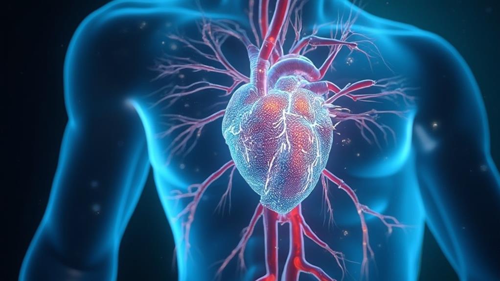
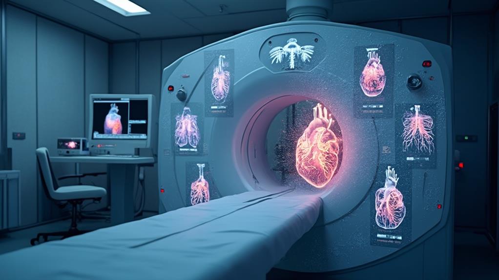
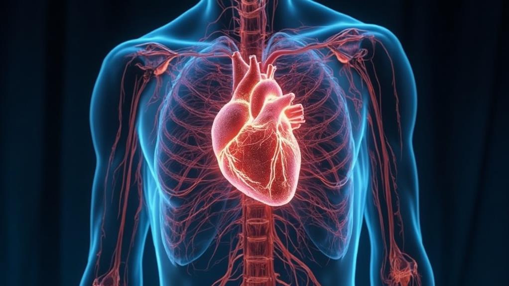
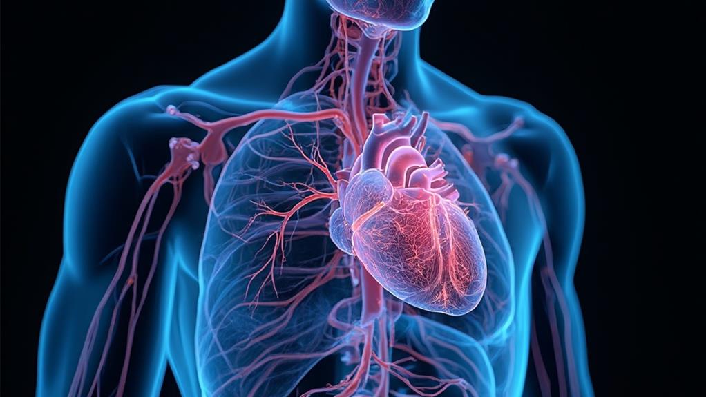
Magnetic Resonance Imaging (MRI) for heart and vascular scans is essential for evaluating cardiovascular health through non-invasive means. Utilizing strong magnetic fields and radio waves, MRI generates detailed images of heart tissues and blood vessels, aiding in the diagnosis and monitoring of conditions such as congenital heart defects, myocardial ischemia, and vascular abnormalities. It provides high-resolution, three-dimensional images without ionizing radiation, which is safer for patients. Limitations include the necessity for patient immobility, high costs, and potential incompatibility with metallic implants. Understanding these aspects can enhance clinical decision-making and improve patient outcomes through advanced imaging techniques.
MRI Highlights
- MRI uses powerful magnets and radio waves to generate detailed images of heart and vascular structures without ionizing radiation.
- MRI angiography (MRA) effectively identifies arterial obstructions, stenoses, and aneurysms in the vascular system.
- MRI aids in diagnosing congenital heart anomalies and evaluating ventricular function such as ejection fraction and wall motion abnormalities.
- MRI provides high-resolution, three-dimensional images, enabling precise visualization of cardiovascular conditions like myocardial infarction and vascular diseases.
- The non-invasive nature of MRI reduces patient discomfort, though it requires immobility and is impacted by the presence of metallic implants.
Definition and Purpose of MRI

Magnetic Resonance Imaging (MRI) operates by using strong magnetic fields and radio waves to generate detailed images of the body's internal structures. In the context of heart and vascular scans, MRI serves critical diagnostic applications by non-invasively evaluating tissue integrity and blood flow.
MRA, a specialized type of MRI that evaluates arteries and blood flow, enhances these diagnostic capabilities by identifying arterial obstructions or narrowing (stenosis). While it offers exceptional benefits such as high resolution and absence of ionizing radiation, it's important to recognize its limitations, including high cost and the need for patient immobility during scans.
How MRI Works
An MRI, or Magnetic Resonance Imaging, is a sophisticated diagnostic tool used to visualize the heart and vascular structures non-invasively. Operating based on magnetic fields and radiofrequency pulses, MRI provides detailed images that assist in diagnosing and monitoring various conditions.
The technology utilizes powerful magnets to align hydrogen protons found in the body's tissues. Once aligned, these protons respond to radiofrequency waves emitted during the scan. When the radiofrequency waves are turned off, the protons return to their normal alignment, emitting signals in the process. These emitted signals are captured and translated by computer algorithms into highly detailed, cross-sectional images.
This method allows clinicians to, without entering the body, observe the heart and surrounding vascular systems with exceptional clarity. Tissues of varying densities and compositions can be distinguished, enhancing the ability to pinpoint irregularities or abnormalities.
The non-invasive nature of MRIs brings comfort and safety to patients, reducing risks associated with more invasive procedures. In addition, MRI avoids exposure to ionizing radiation, making it a safer alternative for repeated imaging. This capability is essential for tracking the progression of diseases and tailoring personalized treatment plans.
Diagnostic Applications
MRI has become an invaluable tool in the domain of diagnostic imaging, particularly for cardiac and vascular evaluations. By leveraging magnetic fields and radiofrequency pulses, Magnetic Resonance Imaging (MRI) provides detailed images of the heart and blood vessels without the need for invasive procedures. This non-invasive technique plays a pivotal role in the early detection and management of various cardiovascular conditions.
Key Diagnostic Applications:
- Congenital Heart Defects: MRI offers detailed visualization of congenital heart anomalies, aiding in accurate diagnosis and treatment planning.
- Cardiac Function Assessment: It allows for precise measurement of ventricular function, including ejection fraction, wall motion abnormalities, and myocardial perfusion.
- Vascular Imaging: MRI angiography (MRA) is exemplary for visualizing blood vessels, identifying stenoses, aneurysms, and other vascular abnormalities.
These applications underscore the importance of MRI in diagnosing complex cardiovascular conditions effectively. Expertise in MRI technology guarantees thorough evaluation and fosters a proactive approach in treating heart and vascular diseases. By providing clear, detailed images, clinicians can better understand the underlying issues, tailoring interventions to optimize patient outcomes and improve quality of life. The role of MRI in cardiac and vascular diagnostics continues to evolve, promising advancements in non-invasive medical imaging.
Benefits and Limitations
Building on the diagnostic capacities highlighted, it is important to understand the broader spectrum of benefits and limitations associated with MRI technology for heart and vascular scans. MRI, or Magnetic Resonance Imaging, leverages powerful magnets and radio waves to create detailed images of the heart and blood vessels without the need for invasive procedures.
This advanced imaging technique excels in providing high-resolution, three-dimensional images that can reveal subtle abnormalities in cardiovascular structures, potentially identifying issues before they manifest as symptoms.
The non-invasive nature of MRI minimizes patient discomfort and eliminates exposure to ionizing radiation, making it a safer option, particularly for repeated use. MRI's versatility extends to evaluating a range of cardiovascular conditions, including congenital heart defects, myocardial infarction, and vascular diseases.
However, limitations exist. MRI requires patients to remain still for extended periods, which can be challenging for some individuals. The presence of metallic implants or devices can also preclude some from undergoing an MRI due to safety concerns. Additionally, the high cost and limited availability in certain regions can restrict access. Balancing these factors is essential for healthcare providers aiming to deliver the best possible care for patients with cardiovascular conditions.
Benefits

Magnetic Resonance Imaging (MRI) for heart and vascular scans offers a range of significant benefits. It provides detailed imaging quality, enabling clinicians to gain extensive insights into cardiac structures and functions without the need for invasive procedures.
In addition, vascular MRI is non-invasive and multi-planar, allowing visualization of blood flow and vessel anatomy, which enhances diagnostic precision. Additionally, MRI facilitates early detection of diseases, supporting timely intervention and thorough heart assessments.
Detailed Imaging Quality
Ever wondered why MRI scans are often hailed as the gold standard in heart and vascular imaging? The detailed imaging quality provided by MRI is unmatched, offering significant insights into anatomical structures and functional aspects of the cardiovascular system. This high-resolution imaging plays a pivotal role in diagnosing and managing heart conditions effectively, benefiting both patients and healthcare providers.
Key advantages of MRI's detailed imaging quality include:
- Superior Anatomical Detail: MRI captures precise images of heart tissues, valves, and blood vessels, allowing for the early detection of abnormalities such as aortic aneurysms, blockages, or congenital defects.
- Functional Imaging: Beyond structural detail, MRI can assess cardiac function. It measures blood flow, quantifies myocardial perfusion, and evaluates ventricular function, giving a holistic view of heart health.
- Tissue Characterization: MRI can distinguish between healthy and damaged tissue, which is vital for identifying conditions like myocardial fibrosis or inflammation, guiding targeted treatment plans.
These benefits exemplify why MRI is an indispensable tool in cardiac care, essential for those dedicated to advancing patient outcomes. By providing unparalleled detail and functional insights, MRI reinforces its position as the premier choice for heart and vascular imaging.
Non-Invasive Procedure
One of the most considerable benefits of MRI technology in heart and vascular scans is its non-invasive nature. Unlike traditional methods requiring catheters or injections, MRI scans eliminate the need for physically intrusive procedures, offering patients a safer and more comfortable experience. The absence of surgical instruments or incisions reduces the risk of infection, complications, and additional recovery time, aligning well with the ethos of patient-centered care.
Furthermore, MRI's non-invasive approach is particularly advantageous for individuals who may be unsuitable for invasive procedures due to health concerns or conditions. It offers a reliable alternative that minimizes the physical and psychological stress often associated with more invasive diagnostic methods. Patients benefit from reduced anxiety and discomfort, fostering a more positive healthcare experience.
Additionally, the non-invasive nature of MRI technology allows for more frequent monitoring of heart and vascular health, facilitating regular follow-ups without the burden of repeated invasive procedures. This capability supports proactive healthcare management, enabling timely interventions that can greatly enhance patient outcomes. In other words, the non-invasive feature of MRI scans embodies a compassionate and efficient approach to diagnosing and monitoring cardiovascular conditions, ensuring the well-being of patients remains at the forefront of medical practice.
Early Disease Detection
Early detection of cardiovascular diseases is a paramount advantage of MRI technology. The non-invasive nature of MRI scans allows for identifying potential issues before they manifest into serious health problems. By detecting conditions such as atherosclerosis, myocarditis, and congenital heart defects early, patients receive timely intervention, which can drastically improve outcomes.
MRI technology excels in providing detailed images that reveal abnormalities undetectable through other diagnostic methods. This precision enables healthcare providers to:
- Identify Early-Stage Disease: Early detection allows for prompt, targeted treatment, reducing the risk of progression to more severe conditions.
- Monitor Disease Progression: Regular MRI scans help track the development of detected abnormalities, aiding in modifying treatment plans as necessary to enhance patient care.
- Guide Preventive Measures: By identifying risk factors and early disease markers, MRI scans support the implementation of lifestyle and medical interventions to prevent the onset of severe cardiovascular conditions.
In this way, MRI technology not only aids in accurate early diagnosis but also supports a proactive approach in cardiovascular health management. As healthcare practitioners committed to serving others, leveraging such advanced diagnostic tools can make a considerable difference in patient outcomes.
Comprehensive Heart Assessment
A thorough heart assessment through MRI technology offers numerous benefits, enhancing the accuracy and breadth of cardiovascular evaluations. MRI scans provide highly detailed images of the heart's structures and function, enabling precise diagnoses and monitoring of conditions such as cardiomyopathy, congenital heart disease, and coronary artery disease.
The non-invasive nature of MRI considerably reduces potential risks associated with traditional methods like cardiac catheterization. This makes it an ideal choice for patients who may be more vulnerable to complications. Moreover, MRI excels in soft tissue characterization, allowing for the differentiation between viable and non-viable heart tissue, which is pivotal for planning appropriate treatments.
MRI's robust imaging capabilities extend to the assessment of blood flow and cardiac perfusion, providing inclusive insights into the heart's performance and identifying issues that might not be visible in other imaging modalities. Notably, the ability to visualize myocardial fibrosis and edema offers early detection and specific interventions, potentially averting progression to more severe conditions.
In facilitating detailed and safe evaluations, MRI technology serves as a central tool for healthcare providers. This enables them to make informed, timely decisions in their mission to improve patient outcomes and offer exemplary cardiovascular care.
Contrast Agent Usage

In the context of MRI for heart and vascular scans, the usage of contrast agents plays a pivotal role in enhancing image clarity, consequently aiding in more accurate diagnoses. There are various types of contrast agents available, each with different compositions and suitability for specific cases. Below is a table summarizing key aspects of these agents:
| Aspect | Details |
|---|---|
| Types of Contrast Agents | Gadolinium-based, Iron-based |
| Safety and Side Effects | Generally safe, rare reactions |
| Enhancing Image Clarity | Improves vessel and tissue detail |
Types of Contrast Agents
Employing contrast agents in MRI for heart and vascular scans considerably enhances the quality of the images obtained, providing more detailed and accurate diagnostic information. Different types of contrast agents are used to achieve this enhancement, each designed to cater to specific diagnostic needs and patient conditions. Generally, these agents contain gadolinium, a heavy metal ion which, when bound to chelating agents, safely facilitates superior image contrast.
There are several categories of contrast agents:
- Extracellular fluid (ECF) agents: Commonly used, ECF agents distribute quickly within the bloodstream and interstitial spaces, enhancing the visibility of vascular structures and tissues.
- Blood pool agents: These agents remain within the vascular system for extended periods, providing prolonged and detailed imaging of blood flow and vessel morphology, beneficial for complex vascular assessments.
- Organ-specific agents: Designed to target specific organs or tissues, these agents attach to structures within the heart or blood vessels, greatly improving the clarity of the targeted region in the MRI scan.
Choosing the appropriate contrast agent maximizes diagnostic efficacy, aiding healthcare providers in making accurate evaluations and improving patient care outcomes.
Safety and Side Effects
When considering the administration of contrast agents in MRI for heart and vascular scans, it is imperative to address their safety and potential side effects to guarantee patient well-being. Contrast agents, often gadolinium-based, enhance the clarity of MRI images, making them indispensable. However, their usage must be meticulously evaluated due to associated risks.
Primary concerns include allergic reactions, although these are relatively rare. Symptoms can range from mild, such as itching and rashes, to severe, including anaphylactic reactions. Screening patients for a history of allergies and prior contrast reactions is critical to mitigate these risks.
Additionally, nephrogenic systemic fibrosis (NSF) is a key condition linked to gadolinium, predominantly affecting those with impaired kidney function. To prevent NSF, patients' renal function is routinely assessed prior to administering gadolinium.
Systemic side effects, though uncommon, also warrant attention. These can include nausea, headaches, or dizziness post-administration. It's essential for medical professionals to remain vigilant and provide appropriate care instructions post-scan. Educating patients about potential side effects and ensuring post-procedure monitoring highlight a commitment to patient safety and extensive care.
Thus, while contrast agents are essential, prioritizing patient safety through stringent screening and monitoring is crucial.
Enhancing Image Clarity
While maintaining patient safety as the top priority, another significant aspect is the enhancement of image clarity through the use of contrast agents. Contrast agents, particularly gadolinium-based compounds, are intravenously administered to improve the differentiation of tissues in MRI scans. This enhancement is particularly indispensable for detailed cardiovascular imaging, allowing clinicians to better discern abnormalities.
Contrast agents function by altering the magnetic properties of nearby hydrogen atoms, thereby increasing the contrast between different types of tissue. This results in clearer, more detailed images, which are essential for accurate diagnosis and treatment planning.
Three primary benefits of using contrast agents in cardiovascular MRI are:
- Improved Visualization: Enhanced clarity helps in identifying finer details of cardiac structures, such as vessel walls and myocardial tissues.
- Better Detection: The enhanced contrast aids in the early detection of pathological conditions such as ischemia, infarction, and fibrosis.
- Increased Diagnostic Confidence: Clearer images allow for more precise measurements and assessments, contributing to more confident and reliable diagnostic decisions.
Ultimately, the use of contrast agents in cardiovascular MRI is a vital tool, facilitating the provision of high-quality care and treatment to patients. This approach ensures clinicians have the best possible information to tailor interventions effectively.
MRI FAQ
How Long Does a Typical Heart and Vascular MRI Scan Take?
A typical heart and vascular MRI scan usually takes approximately 30 to 90 minutes. The duration can vary based on the specific details of the examination and the patient's unique medical conditions. Professional care guarantees ideal results.
Are There Any Specific Preparations Required Before Undergoing an MRI?
Patients should follow specific preparation guidelines such as fasting for a certain period, removing all metal objects, and informing the technician of any implants or allergies. Adhering to these instructions guarantees a smooth and accurate examination process.
What Should I Do if I'm Claustrophobic but Need an MRI?
If you experience claustrophobia, inform your healthcare provider. They may offer a sedative or recommend an open MRI scanner to guarantee your comfort. Communication and preparation are key to a successful and stress-free procedure.
Can I Have an MRI if I Have Metal Implants or Devices?
Patients with metal implants or devices should consult their healthcare provider prior to undergoing an MRI. Certain implants may be compatible, while others could pose risks. This precaution guarantees safety and efficacy in diagnostic imaging.
How Soon Will I Receive the Results of My MRI Scan?
The timeline for receiving MRI results can vary, but typically, the results are available within a few days. Your healthcare provider will review the images and contact you to discuss the findings and any necessary follow-up actions.
