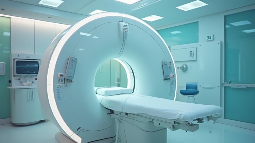
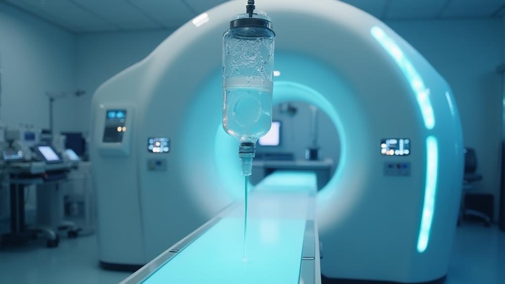
During an MRI, expect to complete a medical history and allergy questionnaire before removing any metallic objects and wearing a hospital gown; a contrast agent may be administered. You will lie on a motorized table, which slides into the MRI bore, and receive earplugs or headphones to mitigate noise. The machine uses powerful magnets and radiofrequency pulses to produce detailed images. Post-scan, you may be monitored for reactions to the contrast dye, and a radiologist will analyze the images, sending the results to your physician. This process enhances diagnostic accuracy and patient safety, providing critical insights into your health.
MRI Highlights
- Patients must remove metallic objects and might receive a contrast dye for enhanced imaging.
- During the scan, patients lie on a motorized table that moves into the MRI machine.
- Earplugs or headphones are provided to reduce the machine’s loud noise.
- The procedure involves magnetic alignment and radiofrequency pulses to produce detailed images.
- Post-scan, patients may need to drink fluids to help expel any administered contrast dye.
MRI Process Overview

What to Expect During an MRI
MRI Process Overview
The MRI process begins with patient preparation steps, including removing metal objects and completing necessary forms. To accommodate patients with specific needs, such as those who are wheelchair-bound or claustrophobic patients, the MRI facility provides a high-field, open MRI design for better comfort.
During the scanning procedure, patients lie on a motorized table that descends for easy access and slides into the MRI machine, where detailed images are captured by utilizing strong magnetic fields and radio waves. After the scan, protocols involve reviewing images for quality and providing post-scan instructions to guarantee patient care is maintained.
Patient Preparation Steps
Preparing for an MRI involves a series of essential steps designed to guarantee patient safety and the accuracy of the imaging results. Initially, patients will be asked to complete a comprehensive questionnaire detailing their medical history, allergies, and any metallic implants or devices within their body. This information helps in identifying potential risks associated with the MRI’s magnetic field.
Next, individuals are instructed to remove all metallic objects, such as jewelry, hairpins, and eyeglasses, to prevent interference with the imaging. For patients requiring a contrast agent for enhanced imaging, the medical staff will explain the nature and purpose of the contrast medium, and an intravenous line may be established.
Prior to the scan, the patient is usually advised to wear a hospital gown to avoid any interactions between regular clothing and the MRI machine. It is also imperative to inform the technologist of any conditions that could affect the scan, such as claustrophobia or pregnancy.
In some cases, pre-scan dietary restrictions are given, like fasting for several hours, especially if sedation or anesthesia is necessary. These steps collectively guarantee the procedure is conducted smoothly, prioritizing the well-being and comfort of the patient.
Scanning Procedure Details
During the MRI scanning procedure, patients are positioned on a motorized table that slides into the cylindrical bore of the MRI machine. The technologist provides earplugs or headphones to mitigate the loud noises generated by the machine. Communication between the patient and technologist is maintained via an intercom system.
Once inside, the MRI machine uses powerful magnets and radio waves to produce detailed images of the internal structures of the body. Key components of the scanning process:
- Magnetic Field Application: The powerful magnetic field aligns the protons in the body’s hydrogen atoms.
- Radiofrequency Pulses: These pulses are then applied, causing the protons to produce signals that the machine detects.
- Data Processing: The MRI system’s computer processes these signals to create highly detailed images.
It is essential for patients to remain still during the scan to guarantee clear and precise images. Each scan can last from several minutes to over an hour depending on the area being examined. Sometimes, contrast agents may be administered intravenously to enhance image quality.
The entire process is non-invasive and typically painless, contributing considerably to diagnosing and planning effective treatments.
Post-Scan Protocols
After completion of the MRI scan, several post-scan protocols guarantee patient safety and diagnostic effectiveness. The first step involves the radiology technologist removing the patient from the MRI machine and assisting them to a comfortable position. This safeguards that any potential dizziness or disorientation from lying still for an extended period is addressed promptly.
Patients who received a contrast agent during the scan are observed for a short period to monitor for any adverse reactions. It is essential to communicate any feelings of nausea, dizziness, or allergic symptoms to the technologist immediately.
The MRI images are then forwarded to a radiologist, a medical professional specializing in interpreting diagnostic images. The radiologist analyses the images thoroughly to identify any abnormalities or areas of concern, ensuring the highest diagnostic quality.
Once the evaluation is complete, a detailed report is prepared and sent to the referring physician. This physician, familiar with the patient’s medical history and baseline condition, discusses the results with the patient at a follow-up appointment, offering an extensive understanding of the findings and potential next steps in treatment.
These protocols ensure patient comfort, accurate diagnosis, and timely communication of results.
Benefits

MRI scans offer numerous benefits, including enhanced diagnostic accuracy and the ability to detect diseases at an early stage. For example, MRI is better for soft tissue imaging, making it a preferred method for examining the brain and spinal cord. As a non-invasive procedure, MRI reduces patient discomfort and eliminates the risks associated with radiation exposure.
These advantages make MRI a valuable tool in modern medicine, contributing to more effective patient care and treatment outcomes.
Enhanced Diagnostic Accuracy
One of the significant benefits of undergoing an MRI is the enhanced diagnostic accuracy it provides. MRIs utilize powerful magnets and radio waves to create detailed cross-sectional images of the body’s internal structures, rendering a more precise diagnosis possible. This non-invasive imaging method is indispensable in identifying abnormalities that other diagnostic tools might miss.
The high-resolution images produced by MRIs allow healthcare professionals to visualize tissues, organs, and skeletal structures with exceptional clarity, facilitating the identification of minute anomalies.
Multiplanar Capabilities: Unlike other imaging techniques, MRI scans can be conducted in multiple planes, such as axial, sagittal, and coronal, offering an extensive view of the area being examined.
Soft Tissue Contrast: MRI is particularly adept at differentiating between various types of soft tissue, making it invaluable in diagnosing conditions related to the brain, spinal cord, joints, and internal organs.
Through enhanced diagnostic accuracy, MRIs assist healthcare providers in devising more effective treatment plans tailored to individual patient needs. This leads to better patient outcomes, ensuring that medical interventions are both timely and appropriate. For anyone committed to serving others, understanding the benefits of MRI is vital in promoting ideal health and well-being.
Non-Invasive Procedure Benefits
While many medical diagnostic procedures necessitate invasive techniques, MRI stands out as a powerful non-invasive alternative, offering significant benefits to patients. An MRI, or magnetic resonance imaging, uses magnetic fields and radio waves to generate detailed images of the body’s internal structures without the need for surgical intervention or exposure to ionizing radiation.
One key advantage of this non-invasive technique is the reduced risk of complications compared to invasive procedures. Patients avoid the potential for infection, bleeding, and other adverse effects associated with procedures that break the skin or require insertion of instruments into the body. Additionally, MRIs are generally painless, which enhances patient comfort and experience.
The lack of ionizing radiation is another critical benefit. Unlike X-rays or CT scans, MRI does not subject patients to radiation, making it a safer option for repeated use, particularly in monitoring chronic conditions or conducting follow-up examinations.
Furthermore, MRIs are suitable for a wide range of patients, including pregnant women and children, who may be more vulnerable to the risks associated with other imaging modalities. The procedure’s non-intrusive nature guarantees that it remains one of the preferred diagnostic tools in modern medicine.
Early Disease Detection
Leveraging advanced imaging technology, early disease detection through MRI provides critical benefits in the clinical setting. MRI, or magnetic resonance imaging, is instrumental in identifying various pathologies at an incipient stage, allowing for more effective interventions. This early detection has the potential to drastically improve patient outcomes, offering a range of advantages for both patients and healthcare providers.
Enhanced Treatment Planning: Detecting diseases early enables healthcare professionals to devise more precise and individualized treatment plans, increasing the likelihood of successful outcomes.
Reduced Healthcare Costs: Early identification of medical conditions can lead to less invasive treatments and shorter recovery times, reducing overall healthcare expenditures.
Improved Prognosis: Early-stage detection often correlates with more favorable prognoses, as many conditions are more treatable when caught early.
In clinical practice, MRI is invaluable for detecting conditions such as tumors, neurological disorders, and cardiovascular diseases long before symptoms manifest. This proactive approach not only enhances patient care but also underscores a commitment to preventive medicine. Through early disease detection, MRI contributes vastly to advancing patient health and enabling timely, targeted therapeutic strategies.
No Radiation Exposure
In an era where minimizing patient exposure to harmful elements is paramount, the distinct advantage of MRI technology lies in its ability to generate detailed internal images without the use of ionizing radiation. Unlike X-rays or CT scans, which employ ionizing radiation to create images of the body’s internal structures, MRI relies on magnetic fields and radio waves.
This makes MRI a safer option for patients who require frequent imaging, such as those with chronic health conditions or those undergoing cancer treatment surveillance.
The absence of ionizing radiation in MRI technology translates to a considerable reduction in potential long-term risks associated with radiation exposure. For particularly vulnerable populations, such as pregnant women, children, and individuals requiring repeat imaging, this advantage cannot be overstated.
MRI offers a non-invasive, risk-free diagnostic tool that healthcare providers can utilize repeatedly without compromising patient welfare.
Moreover, the clarity of the images produced by MRI scans supports accurate diagnoses and informed treatment plans. This combination of precision imaging and enhanced safety guarantees that healthcare practitioners can deliver exceptional care with minimal risk to the patients they serve.
In other words, the no-radiation advantage of MRI exemplifies a commitment to patient-centric, safe medical practices.
Contrast Dye Usage

Contrast dye plays a critical role in enhancing the clarity of MRI images by highlighting specific areas of the body, allowing for improved diagnostic accuracy. While typically safe, patients should be aware of potential side effects, such as allergic reactions or discomfort at the injection site. The following table outlines key aspects related to the purpose, potential side effects, and preparation for contrast dye usage:
| Aspect | Details | Notes |
|---|---|---|
| Purpose | Enhances MRI image clarity | Improves diagnostic accuracy |
| Potential Side Effects | Allergic reactions, discomfort | Generally rare |
| Preparation | Fasting, discussing allergies | Follow medical advice |
| Procedure | Intravenous injection | Monitored by healthcare staff |
| Post-Procedure | Hydration to flush out dye | Medical follow-up if needed |
Purpose of Contrast Dye
The use of contrast dye in an MRI serves a pivotal purpose by enhancing the clarity of the images produced. This special dye, usually injected into the patient’s bloodstream, accentuates specific areas within the body, particularly those that are indispensable for accurate diagnosis. By improving the contrast between different tissues, it helps in distinguishing between healthy and abnormal regions, making it easier for healthcare providers to detect issues such as inflammation, tumors, or blood vessel defects.
In particular, the contrast dye is immensely beneficial in:
- Highlighting vascular structures and blood flow, which aids in diagnosing conditions related to blood vessels.
- Enhancing the visibility of tumors and other abnormal growths, facilitating early detection and precise treatment planning.
- Providing detailed images of soft tissues, including the brain, heart, and digestive organs, ensuring thorough examination.
Administering contrast dye transforms the utility of MRI scans by enabling more precise identification and assessment of medical conditions. This enhancement is pivotal for medical professionals aiming to offer ideal care and effective treatment strategies. For patients, this means receiving more accurate diagnoses and tailored treatment plans that cater specifically to their health needs, ensuring a more informed and targeted approach to their care.
Potential Side Effects
While contrast dye considerably enhances the utility of MRI scans, it is essential to be aware of its potential side effects. The most commonly used contrast agent in MRIs is gadolinium-based. Generally safe, it can rarely cause adverse reactions. Some individuals may experience mild symptoms such as headache, nausea, or dizziness. These side effects usually resolve shortly after the procedure, requiring minimal intervention.
A more serious, though extremely rare, condition is nephrogenic systemic fibrosis (NSF). This primarily affects patients with severe kidney dysfunction. NSF is characterized by thickening and hardening of the skin, along with fibrosis in tissues throughout the body. Consequently, individuals with kidney issues are often pre-screened to mitigate this risk.
For those with allergic reactions to the contrast dye, symptoms can include itching, rash, or hives. Severe anaphylactic reactions are exceedingly rare but necessitate immediate medical attention.
Understanding these potential side effects empowers patients to make informed decisions alongside their healthcare providers. By evaluating individual risk factors, the benefits of contrast-enhanced MRI can often be harnessed safely and effectively. Any concerns or questions should be openly discussed, ensuring patient-centered care and ideal diagnostic outcomes.
Preparation and Procedure
Despite the potential side effects associated with contrast dye, proper preparation can greatly reduce risks and confirm the procedure’s effectiveness. To prepare, patients are often advised to follow specific guidelines that can aid in reducing any adverse reactions and guaranteeing optimal imaging results.
- Medical History Disclosure: Inform the healthcare team about any known allergies, specifically to contrast agents, as well as kidney function concerns.
- Eating and Drinking Instructions: Typically, patients may need to fast for a few hours before the MRI, though clear fluids are generally allowed unless specified otherwise by the healthcare provider.
- Medication Adjustments: Certain medications might need to be paused or adjusted, specifically those affecting kidney function, with advice and instructions coming from your physician.
During the procedure, the contrast dye is usually administered intravenously, and patients might feel a cool sensation as the dye enters the bloodstream. Close monitoring by medical staff ensures any reactions are promptly managed.
After the MRI, patients are often advised to drink plenty of fluids to help flush the contrast dye from their systems. Adherence to these protocols helps ensure the contrast-enhanced MRI yields the most accurate diagnostic images while prioritizing patient safety and comfort.
MRI FAQ
Are There Any Risks Associated With MRI Scans?
While MRI scans are generally safe, there are minimal risks such as allergic reactions to contrast dye, discomfort from loud noises, or issues for individuals with metal implants. Always communicate any health concerns to your healthcare provider.
Do I Need to Fast Before an MRI?
Fasting is generally not required before an MRI, although exceptions exist based on the specific scan type and contrast use. Always follow the guidance provided by your healthcare team to guarantee the most accurate and effective results.
How Should I Dress for My MRI Appointment?
Wear comfortable, loose-fitting clothing free of metallic elements for your MRI appointment. Alternatively, you may be asked to change into a hospital gown. Guarantee all jewelry and metal accessories are removed to guarantee accurate imaging.
Can I Have an MRI if I Have Metal Implants?
Yes, you can undergo an MRI with metal implants, but it requires special considerations. Inform your healthcare provider about any implants, as they will determine the appropriate adjustments or alternative imaging methods to guarantee your safety.
How Long Does an MRI Exam Typically Take?
MRI exams typically take between 30 to 60 minutes, depending on the specifics of the procedure and the areas being examined. It is imperative to remain still to guarantee the highest quality images and accurate diagnostic results.
