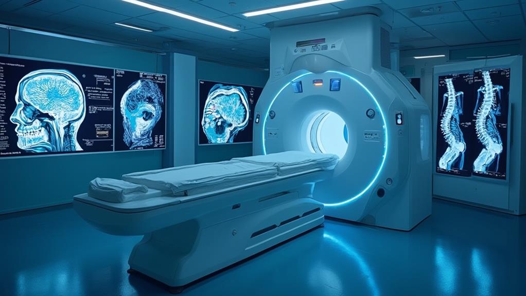
Magnetic Resonance Imaging (MRI) scans are essential in diagnosing and monitoring various medical conditions. Brain and spinal MRIs provide detailed images of neurological structures, crucial for identifying tumors and multiple sclerosis. Cardiac MRIs evaluate heart structures and functions, aiding in the diagnosis of heart diseases and enabling precise visualization of myocardial tissues. Musculoskeletal MRIs focus on bones, joints, and soft tissues, identifying conditions such as arthritis and tendonitis, and tracking post-surgical progress. Each type of MRI utilizes magnetic fields and radio waves to generate high-resolution images. Continuing will offer insights into preparation, benefits, and post-scan guidelines.
MRI Highlights
- Brain and spinal MRI provides detailed images of the nervous system to diagnose tumors, multiple sclerosis, and causes of back pain.
- Cardiac MRI evaluates heart structures and functions, aiding in congenital heart disease, cardiomyopathies, and ischemic heart disease diagnosis.
- Musculoskeletal MRI assesses bones, joints, and soft tissues, helping diagnose arthritis, tendonitis, and bone marrow abnormalities.
- MRI scans use magnetic fields and radio waves for high-resolution imaging without radiation exposure.
- MRI offers three-dimensional reconstructions useful for pre-surgical planning and postoperative evaluations across various medical conditions.
Types of MRI Scans

When considering the various types of MRI scans, it is essential to recognize their specific applications in medical diagnostics.
Brain and spinal MRIs provide detailed images of neurological structures, aiding in the assessment of conditions like tumors and multiple sclerosis. For instance, they can be particularly effective in detecting causes of back pain, such as compression fractures and bone swelling.
Cardiac MRIs focus on heart structures and function, helpful in evaluating congenital heart defects, while musculoskeletal MRIs are indispensable for examining joints, soft tissues, and bone abnormalities.
Brain and Spinal MRI
Magnetic Resonance Imaging (MRI) of the brain and spinal cord is a powerful diagnostic tool widely used in medical practice. This technology utilizes magnetic fields and radio waves to generate detailed images, evaluating healthcare professionals in diagnosing a vast array of neurological conditions.
Brain MRIs can identify abnormalities such as tumors, aneurysms, stroke effects, and multiple sclerosis. These high-resolution images are essential for forming effective treatment plans, improving patient outcomes, and enhancing overall quality of care.
Spinal MRIs, similarly, provide critical information about the spinal cord and surrounding structures. Conditions such as herniated discs, spinal stenosis, and spinal cord injuries can be precisely examined, aiding in timely interventions. The non-invasive nature of MRI makes it an ideal choice, minimizing patient discomfort and avoiding exposure to ionizing radiation.
Furthermore, functional MRI (fMRI) can map brain activity by detecting changes in blood flow, which is instrumental in researching brain disorders and planning surgeries. In both brain and spinal applications, MRI scans are invaluable not just in diagnosing diseases, but also in monitoring disease progression and determining the effectiveness of treatments, ultimately serving the goal of patient-centered care.
Cardiac MRI Applications
Cardiac MRI, an advanced imaging modality, plays a pivotal role in the thorough evaluation of various heart conditions. Utilizing powerful magnetic fields, radio waves, and computer technology, cardiac MRI generates detailed images of heart structures and functions. It is invaluable for diagnosing and managing congenital heart disease, cardiomyopathies, myocarditis, and ischemic heart disease, among others.
One of the primary applications of cardiac MRI is in the assessment and characterization of myocardial infarction, commonly known as heart attacks. It enables precise visualization of scar tissues, aiding in both acute and chronic infarct management. Equally important, cardiac MRI is essential in evaluating myocardial perfusion, revealing regions with impaired blood flow that may require intervention.
Additionally, this imaging modality excels in the detailed visualization of cardiac anatomy, making it particularly useful in the assessment of congenital heart defects. It provides clinicians with three-dimensional reconstructions, vital for pre-surgical planning and postoperative follow-ups.
Cardiac MRI is also instrumental in diagnosing and monitoring cardiomyopathies. It assesses myocardial fibrosis, a key marker in several heart muscle diseases, offering insights into disease progression and therapeutic effectiveness. Hence, cardiac MRI substantially enhances the care and management of patients with diverse cardiac conditions.
Musculoskeletal MRI Uses
Musculoskeletal MRI stands out as an essential tool for diagnosing a wide array of conditions affecting bones, joints, and soft tissues. This imaging modality provides in-depth views of areas like muscles, ligaments, cartilage, and the internal structures of joints. Such precise imaging is pivotal for inspecting injuries that may not be clearly visible through X-rays or CT scans, such as ligament tears, muscle strains, and cartilage degeneration.
Musculoskeletal MRI also plays a critical role in diagnosing chronic conditions. It effectively evaluates disorders such as arthritis, various forms of tendonitis, and even bone marrow abnormalities. The ability to distinguish between the different kinds of tissues enhances its diagnostic accuracy, aiding physicians in formulating tailored treatment plans.
Moreover, musculoskeletal MRI is indispensable in post-operative evaluations. It helps in tracking the progress of surgical interventions and identifying potential complications, ensuring that patients receive timely and appropriate care. Additionally, it is used in cancer detection, identifying tumors in bones or soft tissues with high precision.
Benefits

The benefits of MRI scans are numerous, primarily characterized by their detailed imaging precision, which provides clear and accurate visuals of internal organs and tissues. Additionally, MRI scans are non-invasive, minimizing patient discomfort and eliminating the need for surgical procedures.
The use of high-resolution photos taken by high-field MRI machines guarantees superior image quality, aiding in the accurate diagnosis of medical conditions. These advantages contribute to early disease detection and facilitate the development of personalized treatment plans, enhancing patient outcomes.
Detailed Imaging Precision
Benefitting from advanced technological innovations, detailed imaging precision is one of the cardinal advantages of MRI scans. This sophisticated technique provides clinicians with exquisitely detailed images of internal body structures, enabling accurate diagnosis and effective treatment planning.
The high-resolution images produced by MRI scans capture fine details that often go unnoticed in other imaging modalities like X-rays or CT scans. This capability is imperative in identifying subtle abnormalities in soft tissues, organs, and the nervous system.
Detailed imaging precision is particularly beneficial in areas requiring exact anatomical delineation, such as detecting tumors, identifying abnormalities in the brain and spinal cord, and evaluating joint conditions. This attribute guarantees that healthcare providers can design targeted and personalized treatment regimens, ultimately enhancing patient outcomes. Additionally, the specificity of MRI scans reduces the likelihood of misdiagnosis, contributing to more reliable and efficient patient care.
Furthermore, MRI technology is continually evolving, integrating advancements such as functional MRI (fMRI), which measures and maps brain activity. This precise imaging extends the potential of MRI scans, allowing healthcare providers to make well-informed decisions rooted in comprehensive visualizations of the patient’s anatomy. Consequently, detailed imaging precision in MRI scans plays a pivotal role in improving diagnostic accuracy and patient care.
Non-invasive Procedure
An essential benefit of MRI scans lies in their status as a non-invasive procedure, markedly reducing patient discomfort and risk. Non-invasive techniques such as MRI do not require incisions or the use of instrumentation inside the body, thereby eliminating many of the complications associated with invasive procedures. This aspect is particularly consequential for patients and healthcare providers who place a high value on safety and comfort in medical diagnostics.
The non-invasive nature of MRI scans offers the following benefits:
- Minimal Physical Discomfort: Since no surgery, incisions, or invasive instrumentation is necessary, patients experience minimal physical discomfort during the imaging process. This is imperative, especially for those who might be apprehensive about medical procedures.
- Reduced Risk of Infection: As there is no need to breach the skin or tissues, MRI scans pose negligible infection risks compared to invasive diagnostic techniques. This safety advantage is crucial, particularly in immunocompromised individuals.
- No Recovery Time: Following an MRI scan, patients do not require recovery time or post-procedural care. This facilitates quicker return to daily activities, enhancing overall patient convenience.
- Pain-Free: The entire process is pain-free, allowing patients to remain at ease throughout the examination. This contributes extensively to a positive patient experience.
These advantages underscore the importance of MRI scans as a patient-friendly diagnostic tool.
Early Disease Detection
Early disease detection is one of the paramount benefits of MRI scans, playing a pivotal role in successful medical interventions. With its high-resolution imaging capabilities, MRI can reveal minute anomalies in soft tissues, bones, and organs, often before symptoms manifest. This early detection is critical in diagnosing conditions like cancer, neurological disorders, and cardiovascular diseases, where timely intervention can greatly improve patient outcomes.
Healthcare providers rely on MRI scans to identify subtle changes that other imaging techniques might miss. The ability to detect diseases at an early stage allows clinicians to initiate treatments sooner, potentially halting disease progression and enhancing the efficacy of therapeutic measures. This not only improves the prognosis for patients but also contributes to more efficient healthcare management.
Furthermore, early detection via MRI scans can reduce the need for more invasive diagnostic procedures, sparing patients from the associated risks and discomfort. By providing an extensive view of internal structures, MRIs enable healthcare professionals to make informed decisions about the patient’s condition, fostering a proactive approach to health management. Overall, MRI’s role in early disease detection underscores its invaluable contribution to preventive medicine and patient care.
Personalized Treatment Plans
The exceptional imaging capabilities of MRI scans not only facilitate early disease detection but also empower healthcare providers to formulate personalized treatment plans. These scans deliver detailed images of soft tissues, organs, and other internal structures, allowing for precise diagnosis and tailored therapy approaches.
Enhanced Diagnostic Accuracy: MRI scans provide high-resolution images, which enable healthcare professionals to identify the exact nature and location of a medical issue. This precision is vital for developing a treatment plan that caters to the patient’s specific condition.
Tailored Interventions: With a comprehensive view of the affected area, physicians can design treatment strategies that are specifically tailored to the individual’s needs. This personalized approach can improve patient outcomes and minimize unnecessary treatments.
Monitoring Progress: Individualized treatment plans require ongoing monitoring. MRI scans enable healthcare providers to track the effectiveness of the intervention over time, allowing for adjustments to be made as needed. This continuous evaluation guarantees that the treatment remains effective and aligned with the patient’s recovery.
Improved Patient Engagement: When patients understand the specifics of their condition and the rationale behind their treatment plan, they are more likely to be engaged and compliant with the prescribed regimen. MRI scans help in visualizing the problem, thereby fostering a collaborative relationship between the patient and healthcare provider.
Extra Considerations and Tips

When preparing for an MRI scan, it is vital to reflect on several important factors to guarantee a smooth and effective procedure. Patients should be aware of specific preparation guidelines, appropriate clothing choices, and necessary steps to follow post-scan. The table below outlines key considerations for each stage of the MRI process:
| Aspect | Details |
|---|---|
| Preparation Before Scan | Follow fasting requirements, inform about allergies or health conditions |
| Clothing and Accessories | Wear loose clothing, remove all metal items |
| Post-Scan Guidelines | Await radiologist’s instructions, monitor for any delayed reactions |
Preparation Before Scan
To guarantee an ideal outcome and a seamless experience, thorough preparation before undergoing an MRI scan is indispensable. The relevance of preparation extends beyond just arriving at the appointment on time. Extra considerations and careful planning can enhance comfort and scanning efficiency.
Key preparatory steps to contemplate include:
- Medical History Disclosure: Inform your healthcare provider about any medical conditions, surgeries, or allergies. This information is pivotal, especially if you have implants, such as pacemakers, which could be affected by the MRI’s electromagnetic fields.
- Dietary Restrictions: Follow any provided dietary guidelines. Some scans may require fasting for a specific period. Ensure you are clear about what foods and drinks to avoid, as this can influence scan results.
- Medication Management: Clarify with your doctor whether you should take your regular medications. Certain prescriptions, especially those for blood pressure or heart conditions, may need adjustments prior to the MRI.
- Hydration: Staying well-hydrated is essential. Adequate fluid intake can assist in clearer imaging, especially for scans requiring contrast dye. Drink water as advised by medical staff.
Careful attention to these preparatory steps can substantially contribute to a successful and efficient MRI scanning process.
Clothing and Accessories
Considering clothing and accessories is pivotal for a smooth MRI scanning process. Patients should be mindful of wearing loose-fitting and comfortable garments free from metallic components. Metal objects, including zippers, buttons, and underwire bras, can interfere with the MRI’s magnetic field, leading to distorted images and requiring rescheduling. Clothing provided by the facility may be necessary to comply with safety standards.
Equally important is the removal of all accessories before entering the MRI room. Watches, jewelry, and hairpins must be left outside due to their potential to become projectiles in the magnetic field, posing risks to both the patient and staff. Hearing aids, dentures, and non-permanent retainers should also be removed to prevent interference.
Patients with permanent metal implants, such as pacemakers or aneurysm clips, must inform the MRI technician beforehand. Some implants may be MRI-safe, but others could malfunction, necessitating special protocols.
Hair products containing metallic particles, such as certain sprays or gels, should be avoided. Additionally, tattoos with metallic ink may cause discomfort but rarely pose severe risks; disclosing their presence allows technicians to tailor the scanning approach.
Adhering to these guidelines contributes greatly to a safe and efficient MRI experience.
Post-Scan Guidelines
After completing an MRI scan, adherence to post-scan guidelines guarantees ideal patient recovery and precise diagnostic outcomes. Patients often overlook this important step, which can impact their overall well-being and the accuracy of the diagnostic process.
- Hydrate Well: Drink plenty of water post-scan. This helps in flushing out any contrast material used during the MRI from the body. An increase in fluid intake assists the kidneys in efficiently processing and eliminating these substances.
- Monitor for Side Effects: Pay close attention to any unusual symptoms such as dizziness, headache, or discomfort at the injection site. Although rare, these could signify adverse reactions to contrast materials or other scan-related elements. Report any concerns to healthcare professionals immediately.
- Rest and Recovery: Allocate time to rest, especially if sedatives were administered. Sedation can impair motor skills and judgment, so refrain from activities requiring full alertness such as driving or operating heavy machinery until completely awake.
- Follow-Up Instructions: Adhere strictly to any specific post-scan instructions given by the healthcare provider. This may include taking prescribed medications, attending follow-up appointments, and avoiding certain activities that could interfere with recovery or subsequent tests.
Following these steps guarantees a safe and effective post-scan experience.
MRI FAQ
What Should I Expect During an MRI Scan?
During an MRI scan, expect to lie on a table that moves into the MRI machine. You’ll need to stay still as the machine captures images, experiencing loud noises, but ear protection and communication with technicians guarantee comfort and safety.
Are There Any Risks Associated With MRI Scans?
While MRI scans are generally safe and do not use ionizing radiation, risks include reactions to contrast agents and issues with metallic implants. Always share your medical history to guarantee the safest and most effective care.
How Long Does an MRI Scan Usually Take?
An MRI scan typically ranges from 30 minutes to one hour, depending on the area being examined and the specific type of scan. Patient comfort is prioritized throughout the process to guarantee a positive, service-oriented experience.
Can Anyone Undergo an MRI Scan?
Most individuals can undergo an MRI scan; however, certain conditions like having a pacemaker, metal implants, or severe claustrophobia may necessitate alternative diagnostic methods or special precautions. Consulting a healthcare provider is essential.
Do MRI Scans Require Any Special Preparation?
Yes, MRI scans often require special preparation such as fasting, avoiding certain medications, and removing metal objects. Patients should follow specific instructions provided by their healthcare team to guarantee the procedure’s effectiveness and safety.
