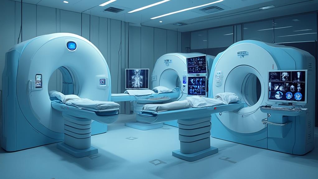
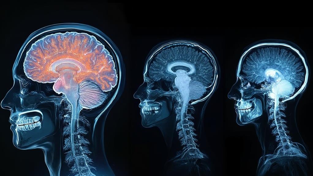
Magnetic Resonance Imaging (MRI) provides superior soft tissue visualization and operates without radiation, making it safer for patients compared to X-rays and CT scans. X-rays offer rapid imaging for structural abnormalities like fractures but lack detailed resolution. CT scans provide highly detailed cross-sectional images using X-ray technology, useful for detecting tumors and guiding surgical procedures. MRI, however, excels in accuracy, especially for complex structures such as the brain and joints, by producing high-resolution images without the long-term risks associated with ionizing radiation. For a deeper understanding of the unique advantages and application specificity of each imaging technique, read on.
MRI Highlights
- MRI provides superior soft tissue visualization without radiation exposure, key for diagnosing complex conditions like brain and joint disorders.
- X-rays offer rapid, cost-effective imaging but lack the detailed resolution needed for soft tissue assessment found in MRI.
- CT scans deliver detailed cross-sectional images, ideal for identifying subtle tissue density differences and planning surgeries, but involve radiation exposure.
- MRI’s non-invasive nature and precision make it safer and more accurate for long-term diagnostic use compared to X-ray and CT imaging.
- MRI helps detect conditions like herniated discs and pinched nerves with higher accuracy, essential for effective treatment planning.
Definition and Significance

Magnetic Resonance Imaging (MRI) stands out for its non-invasive precision in soft tissue visualization, making it invaluable for diagnosing a range of conditions. One notable advantage of MRI is its ability to detect issues like herniated discs and pinched nerves, especially in the spinal cord.
In contrast, X-ray imaging offers quick insight into structural abnormalities like fractures, though it lacks the detailed resolution of other techniques. Computed Tomography (CT) scans provide detailed cross-sectional images, combining the speed of X-rays with enhanced depth, thereby offering a more exhaustive view of internal structures.
MRI: Non-Invasive Precision
MRI stands for Magnetic Resonance Imaging, a technique renowned for its non-invasive precision in medical diagnostics. This sophisticated technology uses powerful magnets, radio waves, and a computer to create detailed images of the body’s internal structures. Unlike other imaging methods, MRI does not involve ionizing radiation, making it a safer option for patients, especially when frequent imaging is required.
The precision of MRI lies in its ability to differentiate between various types of soft tissues. This makes it particularly useful for diagnosing conditions affecting the brain, spinal cord, joints, and internal organs. Medical practitioners can obtain clear, high-resolution images, which are essential for accurate diagnosis and effective treatment planning.
Furthermore, MRI’s non-invasive nature means there is no need for surgical procedures to get a clear view inside the body. This reduces the risk of infection and speeds up the recovery time for patients. It also allows healthcare providers to conduct repeated scans without posing additional health risks.
Ultimately, MRI’s capability to provide detailed, non-invasive imaging plays a pivotal role in modern medical diagnostics, enabling healthcare professionals to serve their patients with greater accuracy and care.
X-Ray: Quick Structural Imaging
X-rays, a form of electromagnetic radiation, are a time-efficient imaging technique extensively utilized in the medical field to visualize the structural integrity of bones and detect abnormalities. This technology is essential for diagnosing fractures, dislocations, infections, and various bone diseases. By providing immediate and clear images, X-rays enable healthcare professionals to make prompt and informed decisions imperative for patient care.
Renowned for its efficiency and utility, X-ray imaging holds several advantages:
- Rapid Results: X-ray procedures typically take just a few minutes, offering near-instantaneous images that facilitate quick diagnostic and therapeutic actions.
- Non-Invasive: As a non-invasive method, X-rays cause minimal discomfort, making them suitable for a broad patient demographic, including those who are critically injured or in pain.
- Cost-Effective: Compared to more complex imaging techniques, X-rays are generally more affordable, making this technology accessible to a larger population and playing a significant role in public health strategies.
- Widespread Availability: X-ray machines are commonly found in hospitals, clinics, and even portable settings, ensuring that almost any medical facility can provide essential imaging services.
These attributes affirm the indispensable role of X-rays in medical diagnostics, aiding in quick, accurate assessments necessary for effective treatment plans.
CT Scan: Detailed Cross-Sections
A CT scan, also known as computed tomography, is an advanced imaging technique that provides highly detailed cross-sectional images of the body’s internal structures. By utilizing a series of X-ray measurements taken from different angles, a CT scan constructs a three-dimensional representation of the targeted area. This method allows for a more extensive view compared to conventional X-rays, which can only offer two-dimensional images.
CT scans are especially valuable in diagnosing and managing various medical conditions. They can detect subtle differences in tissue density, making them particularly useful for identifying abnormalities such as tumors, internal bleeding, and infections. In addition, CT imaging is instrumental in planning and guiding surgical procedures, ensuring precision and safety for patients.
Another significant advantage is the speed at which CT scans can be completed, often providing results within minutes, a pivotal factor in emergency situations.
CT technology has advanced considerably, now including features that reduce radiation exposure while maintaining image quality. This guarantees not only accurate diagnoses but also patient safety. By delivering meticulous cross-sectional views, CT scans remain an indispensable tool in modern medical practice, enhancing healthcare professionals’ ability to serve their patients effectively.
Benefits

One of the key benefits of MRI over other imaging techniques is that it operates without radiation exposure, making it safer for patients.
Additionally, MRI provides superior soft tissue visualization, critical for assessing complex structures like the brain and joints. The high-resolution photos produced by MRI also enable accurate diagnoses, which is particularly important in medical fields that demand precision.
Furthermore, its enhanced diagnostic accuracy and versatile imaging capabilities make it an indispensable tool for a wide range of medical conditions.
No Radiation Exposure
Unlike imaging modalities such as computed tomography (CT) scans and traditional X-rays, magnetic resonance imaging (MRI) stands out due to its lack of ionizing radiation. This absence makes MRI an exceptionally safe choice for patients, eliminating the potential hazards associated with repeated exposure to radiation. Medical professionals often prioritize MRI when cumulative radiation exposure becomes a concern, especially for particular patient populations.
Notable benefits of MRI’s no-radiation feature include:
- Safety for Vulnerable Populations: Pregnant women, children, and individuals requiring frequent imaging can undergo MRIs without risk, critical for minimizing health complications.
- Long-term Health Considerations: MRI’s non-reliance on ionizing radiation reduces the potential long-term risk of radiation-induced conditions, promoting overall patient well-being.
- Repeat Imaging Feasibility: Physicians can order multiple MRI scans for ongoing conditions, aiding in consistent, reliable monitoring without cumulative risk.
- Enhanced Public Health Outcomes: Reduced radiation exposure translates to fewer public health concerns over time, fostering healthier communities.
MRI’s radiation-free nature underscores its utility in diverse clinical scenarios. It enables healthcare professionals to deliver superior care while adhering to principles of safety and efficacy. This advantage aligns with the overarching mission of medical practitioners to optimize health outcomes through thoughtful, patient-centric approaches.
Superior Soft Tissue Visualization
MRI is renowned for its unmatched ability to visualize soft tissues in the human body, making it an indispensable tool in modern diagnostics. This superiority stems from its use of strong magnetic fields and radio waves, which produce detailed images of soft tissues that other imaging techniques, such as X-rays and CT scans, often cannot achieve. Through this advanced technology, physicians are able to obtain clear, high-resolution images of organs, muscles, ligaments, and tendons, providing essential insights that aid in diagnosis and treatment planning.
For conditions involving the brain, spinal cord, and joints, MRI‘s superior soft tissue visualization is particularly advantageous. For instance, it enables the detection of anomalies such as tumors, inflammatory conditions, and degenerative changes with remarkable clarity. Additionally, in cardiovascular imaging, MRI allows detailed visualization of the heart and blood vessels, facilitating the diagnosis of conditions like myocardial infarctions and arteriosclerosis.
Enhanced Diagnostic Accuracy
One of the key benefits of MRI in enhancing diagnostic accuracy includes its ability to:
- Detect early stage diseases: MRI is particularly effective in identifying neurological disorders, tumors, and joint abnormalities in their early stages, which is crucial for timely intervention.
- Differentiate between tissue types: Its superior contrast resolution helps in distinguishing between normal and pathological tissues, aiding in accurate diagnoses.
- Minimize false positives and negatives: The detailed images reduce the likelihood of misdiagnosis, ensuring patients receive appropriate treatment.
- Evaluate treatment efficacy: MRI provides reliable follow-up imaging to monitor disease progression or regression, helping healthcare providers adjust treatment plans accordingly.
Versatile Imaging Capabilities
A hallmark of advanced medical imaging, Magnetic Resonance Imaging (MRI) stands out for its versatile imaging capabilities. It can capture detailed images of almost any part of the body, including the brain, spine, joints, organs, and blood vessels. This extensive ability makes MRI an indispensable tool in diagnosing a wide array of conditions without exposing patients to ionizing radiation, offering a safer alternative to techniques like X-ray and CT scans.
MRI’s versatility extends to functional imaging. This includes functional MRI (fMRI), which maps brain activity by detecting changes in blood flow, and Magnetic Resonance Angiography (MRA), which visualizes blood vessels. These advanced techniques provide insights that are pivotal for pre-surgical planning and evaluating the effectiveness of therapeutic interventions.
Further enhancing its utility, MRI can be tailored using different sequences to highlight specific tissues, enabling the identification of abnormalities with high precision. For example, Diffusion Weighted Imaging (DWI) is sensitive to the movement of water molecules and is particularly useful in detecting early signs of stroke.
The extensive versatility of MRI not only aids in accurate diagnosis but also has a profound impact on patient care, emphasizing its essential role in modern medicine.
Technological Advancements

The technological advancements in medical imaging have revolutionized how diagnostics are performed, greatly enhancing image resolution techniques, accelerating scanning capabilities, and integrating artificial intelligence. These improvements enable faster, more accurate diagnoses, which can notably impact patient outcomes. The following table highlights critical advancements evoking awe and optimism in the field:
| Advancement | Impact | Example |
|---|---|---|
| Enhanced Image Resolution | Greater diagnostic clarity | High-definition MRI scans |
| Faster Scanning Capacities | Reduced wait times | Rapid MRI sequences |
| AI Integration | Improved pattern detection | AI-augmented image analysis |
Enhanced Image Resolution Techniques
Advancing image resolution in medical imaging has fundamentally transformed diagnostic capabilities, directly impacting patient care and treatment planning. High-resolution imaging techniques in MRI and other modalities have enabled unprecedented detail and clarity, facilitating more accurate diagnoses and finer detection of pathological changes.
Modern technological advances have made significant strides in enhancing image resolution in medical imaging devices, bridging the gap between diagnostic precision and patient welfare.
Key advancements include:
- Parallel Imaging Techniques: Utilizes multiple coils to accelerate data acquisition, improving spatial resolution without compromising scan time.
- High-Resolution Detectors: Integration of advanced detectors in modalities like CT and ultrasound increases image clarity, allowing for the visualization of smaller anatomical structures.
- Sub-millimeter Slice Imaging: Techniques in MRI that reduce slice thickness, yielding higher-definition images of tissues and organs, essential for detecting fine pathologies.
- Digital Processing Algorithms: Enhanced graphic processing units (GPUs) and advanced algorithms refine image noise and artifacts, leading to sharper and more precise images.
Improvements in these areas directly translate to better diagnostic outcomes and personalized treatment plans. By continuously innovating and integrating cutting-edge resolution techniques in medical imaging, healthcare professionals are better equipped to serve their patients, fostering advancements that prioritize accuracy and detailed visualization.
Faster Scanning Capabilities
How have technological advancements in medical imaging revolutionized scanning speed and patient throughput? In recent years, significant progress in MRI and other imaging techniques has led to faster scanning capabilities, effectively enhancing patient care.
Modern MRI machines now offer accelerated scan times due to advancements like parallel imaging and compressed sensing. These technologies reduce the duration of each scan, thereby increasing the number of patients that can be accommodated within a given timeframe.
Parallel imaging leverages multiple receiver coils to gather data concurrently, diminishing the required number of phase encoding steps. This results in a substantial reduction in scan times without compromising image quality. Compressed sensing further augments this by reconstructing images from fewer data points, allowing for rapid acquisition while maintaining high resolution.
Moreover, advanced hardware improvements such as stronger magnetic fields and enhanced gradient systems contribute to quicker imaging processes. Increased computational power also plays an indispensable role, enabling real-time data processing and rapid image reconstruction. Consequently, faster scanning capabilities not only improve workflow efficiency but also minimize patient discomfort and reduce wait times.
These technological enhancements underscore our commitment to optimizing patient throughput, ensuring timely and effective diagnosis while maintaining high standards of patient care.
AI Integration in Imaging
Technological strides in medical imaging extend beyond hardware improvements, embracing the domain of artificial intelligence (AI) to further revolutionize diagnostic capabilities. AI integration in imaging enhances accuracy, speed, and the overall quality of diagnosis, which is essential for healthcare professionals aiming to serve their patients effectively.
AI algorithms can analyze vast amounts of imaging data more efficiently than humans, leading to earlier and more accurate detection of anomalies. Here’s how AI benefits medical imaging:
- Improved diagnostic accuracy, reducing the risk of human error.
- Enhanced image quality through sophisticated reconstruction techniques.
- Streamlined workflow processes, saving time for radiologists and technicians.
- Predictive analytics, offering personalized treatment options.
In MRI and other imaging modalities, AI aids in differentiating between benign and malignant lesions with greater precision, potentially leading to better patient outcomes. Additionally, AI-based tools can segment and annotate images automatically, facilitating quicker and more reliable data interpretation. This level of technological advancement translates into higher-quality care by enabling early diagnosis and tailored treatment plans that better serve the diverse needs of patients. As AI continues to evolve, it will certainly play an even more critical role in the future of medical imaging.
MRI FAQ
How Do MRI Costs Compare to Other Imaging Techniques?
MRI costs generally exceed those of other imaging techniques such as X-rays and ultrasound. This is due to the advanced technology and higher operational expenses associated with MRI, reflecting its superior capability in providing detailed anatomical images.
Are There Any Risks or Side Effects of MRI Scans?
MRI scans are generally safe, but patients might experience discomfort due to loud noises, claustrophobia, or slight heating of body tissues. Rarely, they may cause allergic reactions to contrast agents or complications with implanted medical devices.
How Long Does an Average MRI Scan Take?
The duration of an average MRI scan typically ranges from 15 to 90 minutes, depending on the complexity of the examination. Patients are advised to remain still to guarantee accurate results and ideal service to their healthcare providers.
Can MRI Scans Be Performed on Patients With Metal Implants?
MRI scans on patients with metal implants generally require caution, as the strong magnetic field can affect both the imaging quality and patient safety. However, specially designed MRI-compatible implants do exist, allowing safe procedures for affected patients.
What Types of Conditions or Injuries Are Best Diagnosed With MRI?
MRI is highly effective in diagnosing conditions such as brain and spinal cord injuries, soft tissue tumors, ligament and tendon injuries, as well as joint abnormalities. Its precision aids in delivering tailored, patient-centered care.
