Magnetic Resonance Imaging (MRI) is pivotal in cancer diagnosis and management, as it produces detailed, high-resolution images of tumors, allowing for early detection and accurate staging. By evaluating the size, location, and spread of cancers, MRI aids in planning effective treatment and monitoring disease progression. Despite its benefits, MRI accessibility varies due to disparities in infrastructure and financial resources, affecting timely diagnosis and treatment in some regions. Additionally, certain risks, such as the use of gadolinium-based contrast agents, necessitate careful consideration. To understand the extensive role of MRI in enhancing patient outcomes, further exploration into this diagnostic tool is essential.
MRI Highlights
- MRI provides detailed imaging for precise cancer localization and staging.
- MRI enables early detection, leading to timely and effective treatment.
- MRI monitors treatment response and tracks disease progression in cancer patients.
- MRI aids in the accurate planning of surgical and radiation therapy interventions.
- MRI reduces the need for invasive diagnostic procedures, improving patient comfort and safety.
Definition and Purpose of MRI

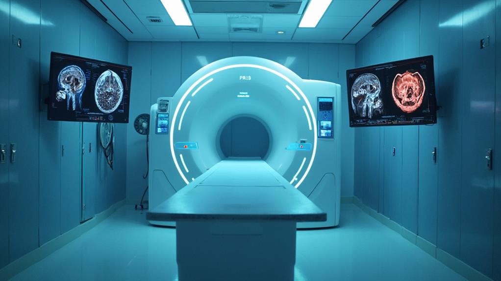
Magnetic Resonance Imaging (MRI) is a sophisticated imaging technology that uses strong magnetic fields and radio waves to generate detailed images of internal structures in the body. It utilizes gadolinium-based contrast agents to improve the visibility of internal structures, enhancing the detection and characterization of various conditions.
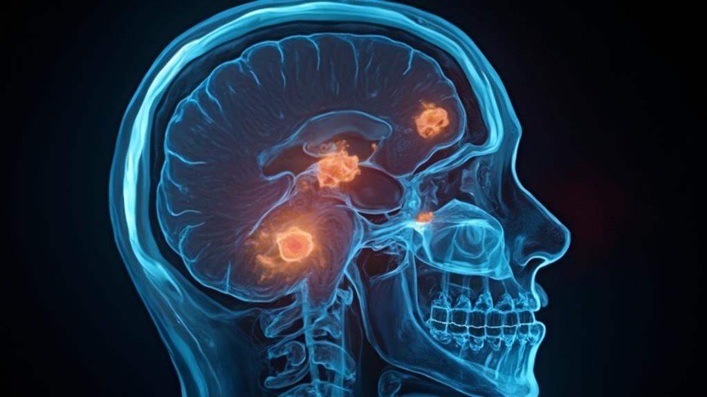
As a non-invasive diagnostic tool, MRI is particularly valuable in cancer diagnosis for its ability to visualize soft tissues with high resolution. By providing extensive and intricate views, MRI aids clinicians in accurately identifying and examining abnormalities such as tumors.
MRI Imaging Technology Basics
Every aspect of modern medicine aims to enhance diagnostic accuracy, and at the forefront of such advancements is Magnetic Resonance Imaging (MRI). MRI uses strong magnetic fields and radio waves to generate detailed images of the internal structures of the body, offering invaluable insights for medical professionals.
Fundamentally, MRI technology involves three key elements:
- Magnetic Field: A powerful magnet aligns the protons in the body's hydrogen atoms. This alignment is indispensable for creating a clear image.
- Radio Waves: Pulses of radio waves are sent through the body, causing these protons to produce signals as they realign with the magnetic field. Each tissue type produces unique signals, allowing for differentiation.
- Computer Processing: These signals are captured by receivers and processed by sophisticated software, constructing highly detailed images that specialists can analyze for abnormalities.
Non-Invasive Diagnostic Tool
As one of the most advanced diagnostic modalities available, MRI stands out for its non-invasive nature and exceptional ability to produce high-resolution images without the need for ionizing radiation. This technology leverages strong magnetic fields and radio waves to generate detailed images of the body's internal structures, making it an invaluable tool in modern medical diagnostics. Remarkably, MRI does not involve the risks associated with ionizing radiation, which is a substantial advantage for patients undergoing repeated scans.
The fundamental purpose of MRI in cancer diagnosis is to provide clear and accurate images that aid in the detection, staging, and monitoring of various malignancies. The non-invasive nature of MRI guarantees that patients are not subjected to unnecessary physical stress or invasive procedures during the diagnostic process. This aligns well with a patient-centered approach, prioritizing comfort and safety while delivering critical diagnostic information.
Furthermore, MRI's ability to differentiate between normal and abnormal tissues enhances its utility in identifying tumors and evaluating their characteristics. The technique's precision contributes significantly to formulating effective treatment plans, thus playing an indispensable role in the comprehensive care of cancer patients. Overall, MRI exemplifies a sophisticated, patient-friendly diagnostic solution.
Detailed Internal Structures Analysis
Uniquely equipped to reveal the complexities of the human body, MRI provides an unparalleled analysis of detailed internal structures. Magnetic Resonance Imaging (MRI) employs strong magnetic fields and radio waves to generate detailed images of organs, tissues, and skeletal structures.
This technique is indispensable in oncology for its ability to create high-resolution images without using ionizing radiation, which is crucial for the accurate diagnosis and staging of cancer.
To further elucidate the value of MRI, consider these key benefits:
- High Resolution and Detailed Imaging: MRI generates images with superior soft tissue contrast, allowing clinicians to distinguish between benign and malignant tissues, assess tumor boundaries, and identify metastasis.
- Multiplanar Capabilities: MRIs can capture images in multiple planes (axial, coronal, and sagittal) without repositioning the patient. This multi-dimensional view aids in the extensive evaluation of irregularly shaped tumors and surrounding structures.
- Functional Imaging Techniques: Advanced MRI techniques, such as diffusion-weighted imaging (DWI) and dynamic contrast-enhanced MRI (DCE-MRI), provide functional and anatomical information. These techniques help in evaluating tumor perfusion, cell density, and response to therapy.
In essence, MRI's detailed analysis of internal structures serves as a cornerstone in the meticulous and compassionate care of cancer patients.
Benefits

MRI technology offers significant benefits in cancer diagnosis, including highly accurate tumor detection and a non-invasive procedure that eliminates the need for surgical interventions.
It is particularly advantageous due to its ability to provide detailed imaging results, which are indispensable for diagnosing many types of medical problems, including cancer. This allows for early identification of cancer, which is vital for effective treatment planning.
Additionally, MRI provides detailed imaging results that help clinicians in creating precise and personalized treatment strategies.
Accurate Tumor Detection
One of the primary benefits of utilizing MRI in cancer diagnosis is its unparalleled accuracy in tumor detection. Magnetic Resonance Imaging (MRI) offers high-resolution images, enabling healthcare providers to detect tumors at their earliest and most treatable stages. This precise imaging capability is critical for forming effective treatment plans and improving patient outcomes.
Additionally, MRI aids in distinguishing between benign and malignant lesions, thereby reducing unnecessary biopsies and surgeries.
Accurate tumor detection through MRI offers several substantial advantages:
- Early Detection: MRI's high sensitivity allows for the identification of very small tumors that might not be visible with other imaging techniques. This early detection can significantly impact the prognosis and treatment success.
- Detailed Imaging: The technology provides detailed images of soft tissues, which is essential for identifying the exact size, shape, and location of the tumor. This detail helps in planning surgical procedures with higher precision, potentially reducing the risk of removing healthy tissue.
- Differential Diagnosis: MRI can differentiate between various types of tissues, which aids in distinguishing between malignant and benign tumors. This differentiation is indispensable for avoiding unnecessary treatments and focusing on malignant cases requiring intervention.
These benefits underscore the pivotal role of MRI in the accurate and efficient detection of tumors, further supporting the mission of healthcare professionals in delivering superior patient care.
Non-Invasive Procedure
Embracing the advantages of non-invasive procedures, the use of MRI in cancer diagnosis streamlines the patient experience while minimizing physical discomfort. Unlike surgical biopsies or exploratory procedures that can carry inherent risks and require recovery time, MRI employs advanced imaging technology to visualize internal structures without the need for incisions or tissue removal. This remarkable benefit allows patients to undergo diagnostics with greater ease and minimal anxiety, promoting a more positive healthcare experience.
MRI's non-invasive nature not only enhances patient comfort but also reduces the potential for complications associated with invasive techniques. The absence of exposure to ionizing radiation further underscores MRI's suitability, as it greatly lowers the risk of radiation-induced side effects, making it a safer option for repeated imaging when necessary. The capability to provide high-contrast images of soft tissues, including tumors, greatly aids healthcare professionals in making accurate diagnoses and formulating precise treatment plans.
For those devoted to providing ideal patient care, the incorporation of MRI as a non-invasive diagnostic tool aligns with the ethical commitment to cause no harm. Overall, the non-invasive benefits of MRI exemplify a superb balance between efficacy and patient-centered care.
Early Cancer Identification
The ability to detect cancer at an early stage substantially enhances the prognosis and treatment options available to patients. Magnetic Resonance Imaging (MRI) plays a pivotal role in this early detection process, providing essential benefits to healthcare professionals and, ultimately, to the patients they serve.
- Improved Prognosis: Early identification of cancer using MRI allows for timely intervention, which is often associated with higher survival rates. Detecting cancer before it spreads significantly increases the chances of successful treatment, reducing mortality rates.
- Less Invasive Treatments: When cancer is diagnosed early, patients can often avoid more aggressive treatments that could be required for advanced stages. This can include smaller surgeries, localized radiation, and potentially fewer or lower doses of chemotherapy, leading to a better quality of life during and after treatment.
- Cost-effective Healthcare: Early diagnosis can lead to reduced healthcare costs over time. Treating cancer in its initial stages often requires fewer resources compared to managing advanced cancer, which may involve complex, prolonged, and more intensive treatments.
Detailed Imaging Results
Incorporating advanced imaging technology, MRI provides remarkably detailed visualizations of internal body structures, which is essential in the accurate diagnosis and management of cancer. High-resolution images allow clinicians to distinguish between different types of tissues, demonstrating variations that might indicate the presence of malignancies. This level of detail facilitates the identification of cancerous tumors, enabling personalized treatment plans tailored to the patient's unique condition.
Moreover, MRI's capacity to provide cross-sectional imaging in multiple planes offers a three-dimensional perspective, which assists in evaluating the size, shape, and exact location of tumors. This is particularly beneficial for planning surgical interventions and radiotherapy, ensuring these treatments are precisely targeted to maximize efficacy while minimizing damage to healthy tissues.
Additionally, MRI does not rely on ionizing radiation, making it a safer imaging option compared to other modalities like CT scans. It is especially advantageous for patients requiring regular follow-ups, thereby reducing cumulative radiation exposure over time. In addition, MRI's exceptional soft-tissue contrast is invaluable for detecting cancer spread to lymph nodes or surrounding structures, guiding inclusive and informed clinical decisions. Embracing MRI in cancer diagnosis embodies a commitment to precision, safety, and a higher standard of patient care.
Limitations and Risks

Despite its benefits, MRI in cancer diagnosis faces certain limitations and risks. Issues such as image resolution constraints, the use of gadolinium-based contrast agents, and limited accessibility pose significant challenges.
| Limitations | Potential Risks |
|---|---|
| Image resolution may be insufficient for detecting very small tumors. | Gadolinium-based agents can cause adverse reactions in some patients. |
| High costs can limit patient access. | Accessibility may be restricted in remote areas. |
| Prolonged scan times may be uncomfortable. | Patients with implants or metal devices may face risks. |
Image Resolution Constraints
One essential challenge in utilizing MRI for cancer diagnosis is the constraint posed by image resolution. This limitation can impact the ability to detect and accurately characterize small or early-stage tumors. High-resolution images are indispensable for a clear and detailed view of anatomical structures, yet the limitations in spatial resolution can lead to several issues.
Tumor Delineation: Lower resolution may prevent precise delineation of tumor boundaries, which is critical for planning surgical interventions or radiotherapy. Blurred edges can obscure the actual size and extent of the tumor.
Lesion Detection: Small lesions may be difficult to detect or may appear indistinguishable from normal tissue. This is particularly problematic in early-stage cancer where prompt detection is key to effective treatment.
Differentiation of Tissue Types: Limited resolution can impede the ability to differentiate between tumor tissue and surrounding normal or benign tissues. This differentiation is essential for accurate diagnosis and treatment planning.
Improving image resolution is a continuous goal in MRI technology to enhance diagnostic capabilities and improve patient outcomes. Ultimately, overcoming these constraints can markedly impact the early and accurate detection of cancer, allowing healthcare professionals to better serve their patients.
Gadolinium-Based Contrast Agents
Gadolinium-based contrast agents (GBCAs) play a pivotal role in enhancing MRI scans to provide better visualization of pathological tissues, including tumors. Despite their indispensable utility in cancer diagnosis, these agents come with limitations and risks that warrant careful consideration by healthcare professionals and patients.
One notable limitation of GBCAs is their nephrotoxicity, particularly in patients with compromised renal function. This can lead to a serious condition termed nephrogenic systemic fibrosis (NSF), characterized by fibrosis of the skin, joints, and internal organs, which can be debilitating. To mitigate this risk, pre-screening for renal function is fundamental before administering GBCAs.
Additionally, while allergic reactions to GBCAs are relatively rare, they can range from mild urticaria to severe anaphylactic reactions. Preparedness to manage these allergic responses is indispensable in clinical settings. Long-term safety concerns also exist, given that gadolinium can deposit in brain tissues even in patients with normal renal function. The clinical implications of such deposits remain under investigation, necessitating continuous monitoring and research.
Limited Accessibility Issues
Although MRI technology has revolutionized cancer diagnosis, its widespread accessibility remains an ongoing challenge, posing significant limitations. One of the primary obstacles is the high cost associated with MRI machines, which can reach upwards of several million dollars, making it impractical for many medical facilities to afford such an investment. This often results in longer wait times for patients in both urban and rural healthcare settings.
Moreover, the availability of trained personnel to operate and interpret MRI scans is another vital barrier. The specialized training required for radiologists and technicians limits the number of qualified professionals, leading to potential delays in diagnosis and treatment. This shortage is exacerbated in underserved areas where healthcare professionals are already scarce.
The inherent limitations include:
- High Operational Costs: Beyond the initial purchase, maintaining MRI machines involves substantial ongoing expenses, including regular maintenance and updates.
- Geographical Disparities: Rural and low-income areas often lack the infrastructure and financial resources to support MRI facilities, widening the healthcare gap.
- Patient Accessibility: Mobility-challenged individuals, particularly those with severe illness, may find it difficult to travel to MRI-equipped centers, impacting early diagnosis and timely treatment.
Addressing these issues is essential to ensuring equitable access to advanced cancer diagnostic tools for all populations.
MRI FAQ
How Should a Patient Prepare for an MRI Scan?
To prepare for an MRI scan, patients should wear comfortable, metal-free clothing, avoid food or drink if instructed, and inform the technician of any medical implants or allergies. Follow specific guidance provided by healthcare professionals.
Can an MRI Detect All Types of Cancer?
An MRI cannot detect all types of cancer; its efficacy varies based on tissue types and cancer characteristics. For essential patient care, it is imperative to integrate MRI with other diagnostic tools for thorough evaluation.
How Long Does an MRI Scan Typically Take?
The duration of an MRI scan typically ranges from 30 to 90 minutes, depending on the area being examined. This variation allows healthcare professionals to deliver precise imaging tailored to patient needs, ensuring thorough diagnostic assessments.
What Is the Cost Range for an MRI Scan in Cancer Diagnosis?
The cost range for an MRI scan typically varies between $400 and $3,500. Variations depend on factors such as location, facility, and insurance coverage. Ensuring accessible pricing is essential for providing thorough care to patients in need.
Are There Any Alternatives to MRI for Cancer Diagnosis?
Yes, alternatives include CT scans, ultrasound, and PET scans. Each method has unique applications and capabilities, offering different benefits based on the type and location of the cancer, as well as patient-specific considerations.
