MRI scans provide detailed imaging essential for diagnosing joint and muscle conditions, offering clear views of muscles, tendons, ligaments, and cartilage. These scans excel in identifying small tears, early-stage strains, and inflammatory conditions like rheumatoid arthritis. MRI is non-invasive, reducing patient risk and discomfort, and safe for repeated use due to the absence of ionizing radiation. Additionally, it assesses cartilage integrity, ligament tears, and meniscal damage, enhancing the quality of musculoskeletal care. Though generally safe, contrast agents used in some MRIs may cause allergic reactions, making thorough patient history assessment indispensable to tailor treatment plans effectively.
MRI Highlights
- MRI provides detailed imaging of muscles, tendons, ligaments, and cartilage, aiding in diagnosing soft tissue injuries.
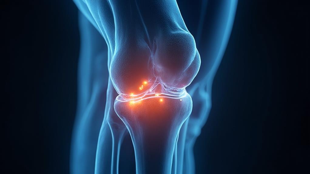
- MRI can visualize joint structures, assessing conditions like meniscal tears and ligament strains.
- MRI detects inflammatory conditions, such as rheumatoid arthritis and bursitis, through subtle imaging changes.
- MRI offers clear, non-invasive imaging without radiation, ensuring patient safety and comfort.
- MRI can identify minute tears and early-stage strains, facilitating timely and targeted interventions.
MRI Purpose Defined

MRI scans are valuable diagnostic tools for evaluating a variety of joint and muscle conditions. They are essential for diagnosing soft tissue injuries, detecting inflammatory conditions, and visualizing joint structures. This non-invasive imaging technology enhances the accuracy of medical evaluations and treatment plans.
MRI is particularly useful for diagnosing sports-related injuries and can provide detailed images of structures that are difficult to image with other modalities.
Diagnosing Soft Tissue Injuries
When evaluating soft tissue injuries, how effectively can modern imaging techniques identify the root causes of pain and dysfunction? Magnetic resonance imaging (MRI) excels in providing detailed images of the body's soft tissues, such as muscles, tendons, ligaments, and cartilage. It does so through the use of powerful magnets and radio waves, which create high-resolution images differentiating between various tissue types, allowing for accurate diagnosis and effective treatment plans.
MRIs are particularly invaluable when dealing with complex injuries that might not be visible through X-rays or ultrasounds. For instance, they can reveal minute tears in ligaments or early-stage muscle strains, conditions often responsible for persistent discomfort. This capability is essential for healthcare professionals dedicated to delivering precise and compassionate care, as it facilitates targeted interventions that address the specific injury.
Furthermore, MRIs are non-invasive, avoiding the risks associated with surgical diagnostic methods. This approach not only reduces patient anxiety and discomfort but also expedites the diagnostic process, helping healthcare providers offer timely and appropriate responses to injuries. In this way, MRI technology enhances the capacity of medical professionals to deliver high-quality, efficacious care to individuals suffering from soft tissue injuries.
Detecting Inflammatory Conditions
Addressing the complexities of soft tissue injuries with advanced imaging techniques naturally leads to the broader utility of MRI in detecting inflammatory conditions. Magnetic Resonance Imaging provides a powerful tool for identifying and evaluating a range of inflammatory diseases impacting joints and muscles. By leveraging its high-resolution imaging capabilities, MRI can detect even subtle changes associated with inflammation, such as synovitis and tenosynovitis, which might not be visible on other imaging modalities like X-rays or CT scans.
The effectiveness of MRI in detecting inflammation is paramount in diagnosing conditions such as rheumatoid arthritis, bursitis, and myositis. Its ability to visualize both soft tissues and bone marrow changes helps medical professionals develop accurate and tailored treatment plans. Additionally, MRI's non-invasive nature and lack of ionizing radiation make it a safe option for repeated use, providing ongoing monitoring of inflammatory conditions over time.
Through enhancing diagnostic precision, MRI supports healthcare providers in making informed decisions that benefit patient care. This technological advancement underscores MRI's essential role in managing inflammatory conditions, emphasizing its importance in achieving timely, accurate diagnoses and ultimately improving patient outcomes.
Visualizing Joint Structures
Understanding the intricate structures within joints is essential for diagnosing and treating a multitude of musculoskeletal conditions. Magnetic Resonance Imaging (MRI) offers unparalleled visualization of these complex areas, making it invaluable for healthcare professionals. Unlike X-rays or CT scans, MRI provides detailed images of soft tissues, cartilage, and ligaments, elucidating pathologies that might not be visible through other imaging modalities.
MRI is particularly adept at:
- Assessing Cartilage: It visualizes cartilage thickness and integrity, vital for diagnosing conditions like osteoarthritis.
- Evaluating Ligaments: For identifying tears or strains in ligaments that X-rays might miss, such as those in the knee or shoulder.
- Visualizing Menisci: Fundamental for diagnosing meniscal tears, often seen in athletes or aging populations.
- Detecting Synovial Fluid Changes: Helps in identifying inflammation or infections within the joints.
These capabilities ensure that MRI scans enable healthcare providers to deliver more precise and extensive care. By directly visualizing key structures, physicians can devise more accurate treatment plans tailored to the needs of each patient. Embracing such advanced imaging techniques is fundamental in enhancing the quality and effectiveness of musculoskeletal healthcare, ultimately fostering better health outcomes for those served.
Benefits

One of the primary benefits of MRI for joint and muscle scans is its ability to provide accurate diagnostic imaging without the need for invasive procedures. This technology is particularly effective for detailed soft tissue analysis, allowing healthcare providers to detect abnormalities at an early stage.
Additionally, MRI facilitates early disease detection, which can noticeably improve patient outcomes by enabling prompt and appropriate treatment interventions. With imaging muscles and joint problems such as arthritis, it becomes easier to diagnose and handle sports-related injuries efficiently.
Accurate Diagnostic Imaging
Accurate diagnostic imaging through MRI offers numerous benefits for joint and muscle scans, considerably enhancing the ability to diagnose and manage musculoskeletal disorders. Magnetic Resonance Imaging (MRI) provides clear and detailed images of soft tissues, such as muscles, tendons, and ligaments, which are essential for identifying injuries and abnormalities that may not be visible through other diagnostic methods.
MRI's ability to distinguish between different types of soft tissue allows for more precise evaluations, which is particularly useful in the detection of tears, inflammation, and degeneration. This accuracy is indispensable for tailoring effective treatment plans and monitoring the progression or improvement of a condition.
Key benefits of accurate diagnostic imaging through MRI include:
- Detailed Visualization: MRI produces high-resolution images, enabling the identification of subtle changes in soft tissues.
- Early Detection: Early and precise diagnosis can lead to more effective and timely treatment interventions.
- Comprehensive Assessment: MRI can evaluate multiple structures in a single scan, offering a holistic view of the affected area.
- Avoidance of Exploratory Surgery: Detailed imaging can reduce the need for invasive diagnostic procedures, minimizing patient risk and discomfort.
These advantages affirm MRI's pivotal role in the healthcare field, ensuring precise diagnosis and ideal patient care in musculoskeletal health.
Non-Invasive Procedure
The non-invasiveness of MRI presents considerable benefits, making it a preferred choice for joint and muscle scans. One of the most prominent advantages is patient comfort. Unlike surgical or invasive diagnostic procedures, MRI scans do not involve incisions or injections into the muscles or joints. This eliminates the associated risks of infection, bleeding, and extended recovery times.
Additionally, the lack of radiation exposure in MRI scans makes it a safer option compared to X-rays or CT scans. This is especially beneficial for patients who require multiple follow-up exams over time. MRI's safety profile reassures patients concerned about the cumulative effects of radiation.
The procedure is also highly precise, aiding healthcare professionals in diagnosing conditions without the need for more intrusive methods. This accuracy can lead to earlier detection and treatment, improving patient outcomes substantially. Furthermore, MRIs can be safely used for a broad demographic, including children and pregnant women, further demonstrating its versatility.
Ultimately, the non-invasive nature of MRIs simplifies the patient experience and aligns with modern healthcare's goal to provide effective, compassionate care. This makes MRI an invaluable tool in the domain of joint and muscle diagnostics.
Detailed Soft Tissue Analysis
Advanced MRI technology provides exceptional capability for detailed soft tissue analysis, which is essential in diagnosing and treating joint and muscle conditions. MRI scans offer unparalleled resolution, allowing healthcare professionals to visualize intricate structures within muscles, tendons, ligaments, and cartilage. This high level of detail is crucial for accurate diagnoses and effective treatment planning.
The benefits of detailed soft tissue analysis using MRI include:
- Enhanced Image Clarity: MRIs produce high-contrast images that vividly display soft tissues, providing clear delineations that help identify abnormalities with precision.
- Non-Invasive Insight: Unlike exploratory surgery, MRI scans offer a non-invasive way to assess internal structures, reducing patient discomfort and associated risks.
- Comprehensive Tissue Evaluation: MRIs facilitate the thorough examination of soft tissue integrity and functionality, aiding in identifying tears, inflammations, and other pathologies.
- Personalized Treatment Plans: Detailed imaging allows for tailored treatment strategies, ensuring interventions are specifically targeted to the individual's unique condition.
These capabilities empower medical professionals to better serve their patients, enhancing the standard of care. Precise soft tissue visualization aids in early and accurate diagnoses, ultimately contributing to improved patient outcomes. As a cornerstone in contemporary medical diagnostics, MRI continues to revolutionize how joint and muscle conditions are understood and treated.
Early Disease Detection
Through early detection of diseases, MRI technology offers significant benefits in the domain of joint and muscle health. Magnetic Resonance Imaging (MRI) excels at identifying conditions in their nascent stages, which is critical for successful intervention and treatment.
Early diagnosis through MRI can reveal subtle changes in soft tissues, cartilage, and bone structures that might not be visible through other imaging techniques like X-rays or CT scans. This early identification empowers healthcare providers to design personalized and timely treatment plans, potentially preventing disease progression.
For patients, the advantages are manifold. MRI scans can detect early signs of conditions such as arthritis, tendonitis, or even muscular dystrophy, enabling interventions that can reduce pain, improve mobility, and enhance quality of life.
In addition, the non-invasive nature of MRIs ensures that patients undergo minimal discomfort during the diagnostic process, making it an appealing option for those who may be apprehensive about more invasive procedures.
Contrast Material Usage

Using contrast material in MRI scans of joints and muscles greatly enhances image clarity, allowing for the precise identification of abnormalities. The procedure involves injecting a contrast agent, usually gadolinium-based, into the patient's bloodstream to improve the differentiation of tissues. However, it is important to be aware of potential side effects, such as allergic reactions or nephrogenic systemic fibrosis.
| Aspect | Details |
|---|---|
| Benefits of Contrast | Enhanced image clarity for better diagnosis |
| Application Procedure | Injection of gadolinium-based agent into bloodstream |
| Potential Side Effects | Allergic reactions, nephrogenic systemic fibrosis |
| Monitoring and Safety | Pre-screening for kidney issues, monitoring during scan |
Benefits of Contrast Material
Contrast material, often employed in magnetic resonance imaging (MRI), offers pivotal benefits that enhance diagnostic accuracy. It plays a vital role, particularly in scans involving joints and muscles. By improving the visibility of specific anatomical structures, contrast material helps healthcare professionals make more precise diagnoses, ensuring patients receive the most suitable treatments.
Key benefits of using contrast material in MRI include:
- Improved Image Contrast: Contrast agents accentuate the differences between various tissues, making it easier to distinguish between normal and abnormal areas.
- Enhanced Detection of Pathologies: Subtle abnormalities, including inflammation, infection, and small lesions, are more easily identified with contrast material, leading to timely intervention.
- Better Vascular Imaging: For musculoskeletal issues involving blood vessels, contrast material helps highlight vascular structures, aiding in the assessment of vascular anomalies or injuries.
- More Detailed Tumor Visualization: When evaluating potential tumors in joints and muscles, contrast material can delineate tumor boundaries more clearly, which is indispensable for treatment planning.
The use of contrast material in MRI substantially elevates the diagnostic capability, providing detailed and reliable images essential for accurate assessment. Consequently, it facilitates effective patient care, ensuring individuals receive the correct diagnosis and appropriate management for their conditions.
Application Procedure Overview
Administering contrast material in MRI procedures involves a series of well-coordinated steps designed to guarantee patient safety and image quality. These steps begin with a comprehensive patient assessment, including reviewing medical history and potential allergies. The healthcare professional explains the procedure, addressing any patient questions to confirm they understand the process and purpose of the contrast material, which enhances tissue differentiation.
The contrast material, typically gadolinium-based, is administered intravenously. The dosage depends on the specific imaging requirements and the patient's body weight. Proper sterilization is vital to prevent infection, and the injection site is carefully selected and cleaned.
Once the contrast material is injected, it circulates through the bloodstream, allowing it to highlight abnormalities in joints and muscles more effectively. Throughout the procedure, the patient is monitored for any immediate reactions. It is pivotal to maintain clear communication and provide reassurance. The MRI scan commences after the contrast material has been administered, capturing detailed images that aid in accurate diagnosis. This careful and systematic approach ensures that the use of contrast material in MRI scans upholds the highest standards of patient care and diagnostic precision.
Potential Side Effects
While the administration of contrast material in MRI scans offers significant diagnostic benefits, it is important to ponder the potential side effects that may arise. Contrast agents, particularly gadolinium-based ones, are widely used to enhance image quality, providing indispensable insights into joint and muscle conditions. However, their use is not without risks.
Common adverse effects associated with contrast material usage include:
- Allergic Reactions: Some patients may experience mild to moderate allergic reactions, such as itching, rashes, or hives.
- Nephrogenic Systemic Fibrosis (NSF): Though rare, NSF is a serious condition that can occur in patients with impaired kidney function when exposed to gadolinium-based contrast agents.
- Metallic Taste: A transient metallic taste is often reported during or shortly after the injection of the contrast material.
- Nausea and Vomiting: Some individuals may feel nauseated or may vomit shortly after the administration of the contrast agent.
Professionals performing MRI scans must assess patient history extensively, particularly for allergies or kidney issues, to mitigate these risks. By doing so, they can guarantee that the diagnostic benefits of the MRI scan are maximized while providing safe and compassionate care to their patients.
MRI FAQ
How Long Does an MRI Scan for Joints and Muscles Typically Take?
The duration of a scan typically ranges from 30 to 60 minutes. The exact time depends on the specific area being examined and the complexity of the scan. Your patience guarantees accurate diagnostics, aiding effective treatment.
Are There Any Risks Associated With MRI Scans for Joint and Muscle Evaluation?
The risks associated with MRI scans for joint and muscle evaluation are minimal, primarily involving possible discomfort from lying still, potential claustrophobia, and rare allergic reactions to contrast agents, thereby ensuring a generally safe diagnostic procedure.
Can I Eat or Drink Before Undergoing a Joint or Muscle MRI?
Patients may generally eat and drink before undergoing a scan unless specific instructions are provided otherwise. However, it is always advisable to follow any preparatory guidelines given by the healthcare provider to guarantee accurate results.
What Should I Wear for an MRI Scan of My Joints or Muscles?
When undergoing such a scan, wear loose-fitting, comfortable clothing without metal components. Alternatively, facility-provided gowns are available. Removing jewelry and metallic items is critical to guarantee image clarity and personal safety during the procedure.
How Should I Prepare for My Joint or Muscle MRI Appointment?
To prepare for your appointment, avoid wearing jewelry and clothing with metal components. Inform the technician about any implants or medical conditions. Arrive on time, follow fasting instructions if provided, and maintain clear communication.
