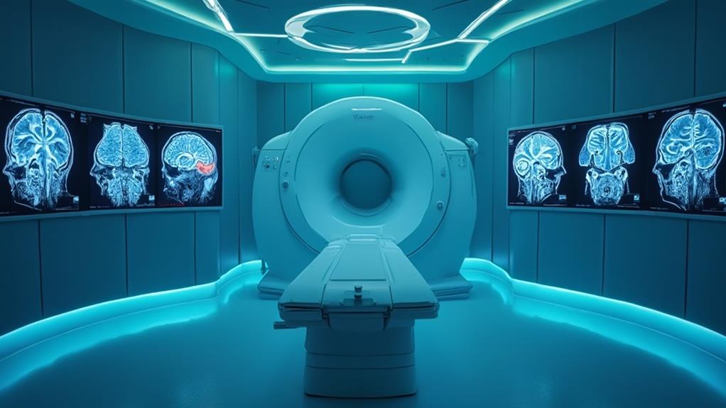
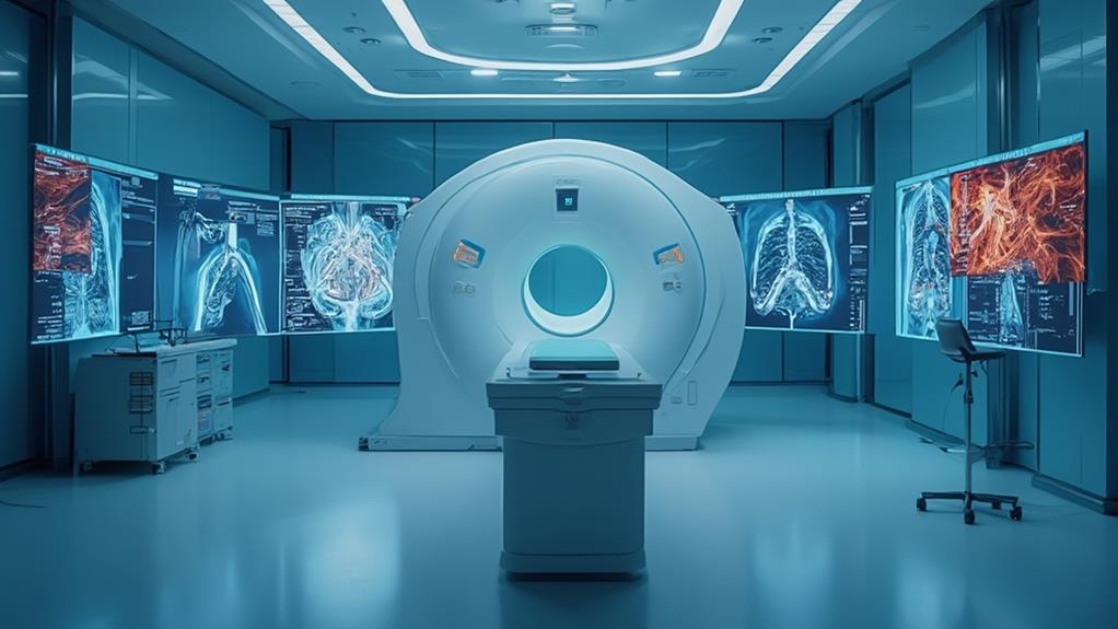
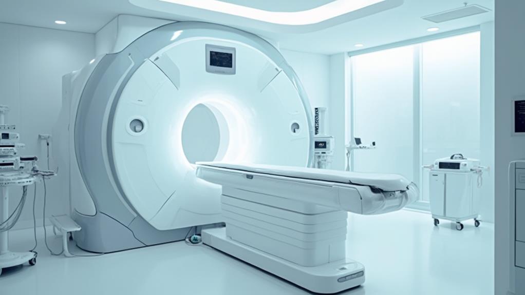
Magnetic Resonance Imaging (MRI) is a sophisticated, non-invasive diagnostic tool that leverages magnetic fields and radio waves to generate thorough images of the body's internal structures and pathologies. It excels in providing high-resolution pictures of soft tissues, making it invaluable for diagnosing neurological, musculoskeletal, and cardiovascular conditions. MRI is particularly adept at differentiating between benign and malignant tumors, detecting early-stage abnormalities, and offering a safe alternative to ionizing radiation. However, potential concerns include claustrophobia, lengthy scan times, and contraindications related to metal implants. Ensuring patient safety and comfort is paramount, as continued advancements reveal further diagnostic potential.
MRI Highlights
- MRI generates detailed images using magnetic fields, enabling non-invasive visualization of internal structures and pathologies.
- It provides superior contrast resolution between soft tissues, ideal for diagnosing neurological, musculoskeletal, and cardiovascular disorders.
- High-contrast imaging capabilities make MRI adept at detecting small or early-stage tumors and complex lesions.
- MRI is a safer diagnostic option as it avoids ionizing radiation and reduces the need for invasive methods.
- Early disease detection through MRI facilitates timely interventions, improving patient outcomes and reducing healthcare costs.
Definition and Function of MRI

Magnetic Resonance Imaging (MRI) serves as a non-invasive diagnostic tool, offering precise imaging of soft tissues, which is essential for identifying anomalies such as tumors and lesions. Its ability to produce detailed images makes it indispensable in detecting and monitoring various medical conditions.
Imaging Soft Tissues Precisely
In the domain of medical diagnostics, MRI stands as a pivotal technology, allowing for high-resolution imaging of soft tissues with remarkable accuracy. By utilizing strong magnetic fields and radio waves, MRI captures detailed images of organs and structures within the body, providing invaluable information for healthcare professionals.
MRI excels in soft tissue imaging due to its ability to differentiate between various types of tissues with high precision. This makes it indispensable in evaluating conditions that affect the brain, spine, joints, and other essential areas. The primary factors that contribute to its effectiveness include:
- Superior contrast resolution: MRI provides excellent contrast between different soft tissues, surpassing the capabilities of other imaging modalities like X-rays and CT scans.
- Non-invasive technique: MRI does not involve exposure to ionizing radiation, making it a safer option for repeated imaging.
- Multiple plane imaging: MRI can generate images in multiple planes (axial, sagittal, coronal), allowing for thorough evaluation from various angles.
These capabilities ensure that MRI is not only a cornerstone in diagnosis but also in the ongoing monitoring and assessment of treatment efficacy. As a result, MRI plays a pivotal role in delivering high-quality care tailored to individual patient needs.
Detecting Tumors and Lesions
Magnetic Resonance Imaging (MRI) is a pivotal technology in medical diagnostics, known for its unparalleled ability to visualize both normal and abnormal tissue structures within the body. Specifically, MRI excels in detecting tumors and lesions, due to its high-contrast imaging capabilities.
Utilizing powerful magnets and radio waves, MRI produces detailed images of the body's internal structures, enabling healthcare professionals to identify abnormalities that may not be visible with other imaging modalities, such as X-rays or ultrasounds.
The advanced technology allows for the differentiation between benign and malignant tumors with a high degree of accuracy. MRI scans can capture variations in tissue density and composition, assisting in the identification of small or early-stage tumors that might otherwise be overlooked.
Additionally, the technique is particularly adept at imaging soft tissues, such as the brain, liver, and spinal cord, where lesions are often complex and difficult to detect.
Non-Invasive Diagnostic Tool
Offering a crucial and non-invasive approach, MRI functions as an essential diagnostic tool by leveraging magnetic fields and radiofrequency pulses to generate detailed images of the body's internal structures. This technology stands out for its ability to produce high-resolution images without the need for surgical procedures or exposure to ionizing radiation, making it a safer option for patients.
The operation of an MRI involves several key components:
- Magnet: A strong magnetic field aligns the protons in the body's hydrogen atoms.
- Radiofrequency Coils: These coils send pulses that knock the protons out of alignment, causing them to emit signals.
- Computer System: Advanced software converts these signals into detailed images of soft tissues, organs, and other internal structures.
MRI is particularly beneficial for diagnosing a range of conditions such as neurological disorders, musculoskeletal injuries, and cardiovascular diseases. It offers superior contrast between different tissues, thus helping healthcare professionals in identifying and examining anomalies with precision. The non-invasive nature of MRI not only enhances patient comfort but also reduces risks associated with invasive diagnostic methods, ensuring a more reliable and patient-centered diagnostic experience.
Benefits

MRI offers several significant advantages, including its status as a non-invasive imaging technique that allows for detailed visualization of soft tissues. This capability is vital for early disease detection, providing critical information without the use of ionizing radiation.
MRI scans can analyze ligament, tendon, and muscle tears, offering clear insight into sports-related injuries. Consequently, MRI is a safer option for repeated imaging, especially when monitoring chronic conditions.
Non-invasive Imaging Technique
Magnetic Resonance Imaging (MRI) stands as a powerful diagnostic tool due to its non-invasive nature, offering significant benefits in medical imaging. Its ability to provide detailed internal images without the need for incisions or exposure to ionizing radiation makes it a preferred method for diagnosing a variety of conditions safely and effectively.
MRI uses magnetic fields and radio waves instead of ionizing radiation, which means it poses no significant health risks to patients, rendering it suitable for repeated use without adverse effects. This characteristic is particularly beneficial for patients requiring long-term monitoring.
The absence of invasive procedures reduces the risk of complications, such as infections or adverse reactions to contrast agents. By eliminating physical intrusion, MRI minimizes discomfort and provides an accurate depiction of the body's internal structures without the potential hazards associated with invasive techniques.
As a non-invasive procedure, MRI facilitates a less stressful experience for patients. The process generally involves lying still inside the MRI machine, a far less intimidating prospect than surgical interventions. This aspect is particularly important for pediatric and elderly patients who may have heightened sensitivity to invasive procedures.
These benefits underscore why MRI is a cornerstone in modern diagnostic medicine, continuously enhancing patient care.
Detailed Soft Tissue Visualization
One of the paramount advantages of MRI technology is its unparalleled capacity for detailed soft tissue visualization. This feature is indispensable in diagnosing a wide array of medical conditions, particularly those involving muscles, tendons, ligaments, and other soft tissues. MRI scans produce highly detailed images, allowing medical professionals to pinpoint abnormalities with exceptional accuracy.
The ability to visualize soft tissues in such detail enhances the diagnostic process, enabling the identification of issues that may not be visible through other imaging modalities, such as X-rays or CT scans. This is especially beneficial when examining complex structures within the body, such as the brain, spinal cord, and joints, where precise imaging is critical for effective diagnosis and treatment planning.
Moreover, detailed soft tissue visualization through MRI aids in evaluating the extent of injuries, such as muscle tears or ligament damage. This information is essential for devising accurate treatment plans and monitoring the healing process. For patients, this means a higher likelihood of receiving the correct diagnosis and appropriate care, ultimately leading to better health outcomes. Overall, the detailed images produced by MRI technology play a pivotal role in advancing patient care and improving the efficacy of medical interventions.
Early Disease Detection
Early detection of diseases offers a substantial advantage in medical diagnosis and treatment, and advanced imaging technologies contribute immensely to this endeavor. Magnetic Resonance Imaging (MRI) plays a pivotal role in identifying conditions at their inception, providing a nuanced view of a patient's internal structures. This capability facilitates early intervention, potentially altering the disease's trajectory in a favorable manner.
The benefits of MRI in early disease detection include:
- Improved Prognosis: Identifying diseases like cancer in their early stages dramatically enhances the chances of successful treatment and long-term survival. For example, MRI can detect small, early-stage tumors that might be missed by other imaging techniques.
- Personalized Treatment Plans: Early detection allows healthcare providers to tailor treatment strategies to the patient's specific condition, leading to more effective and targeted therapies. This individualized approach guarantees that patients receive the most appropriate care based on their unique medical needs.
- Reduced Healthcare Costs: Diagnosing diseases early can reduce the overall cost of healthcare by preventing the progression of the disease to more advanced and costly stages. Early detection and intervention often require less intensive and less expensive treatments.
No Radiation Exposure
A significant benefit of MRI technology is the absence of ionizing radiation, which makes it a safer option for patients. Unlike X-rays and CT scans, MRI uses powerful magnets and radio waves to create detailed images of the body's internal structures. This method eliminates the risk of radiation exposure, which can be particularly harmful with repeated use.
For patients requiring frequent imaging, such as those with chronic conditions like multiple sclerosis or joint disorders, the non-radiative nature of MRI becomes profoundly advantageous. It's also reassuring for parents and healthcare providers when imaging is required for children, as the growing bodies of young patients are more susceptible to the harmful effects of ionizing radiation.
Moreover, the non-use of radiation makes MRI a preferred choice for sensitive areas, such as the brain, spinal cord, and soft tissues, where precision is critical and the avoidance of radiation is indispensable.
The absence of ionizing radiation in MRI not only safeguards the immediate safety of the patient but also contributes to long-term health, reducing the cumulative risks associated with radiation exposure over time. Consequently, MRI emerges as a conscientious choice in diagnostic imaging for maintaining patient safety and health.
Limitations and Safety Concerns

Despite the numerous advantages of MRI in diagnosing various conditions, it is important to acknowledge some limitations and safety concerns associated with its use. Issues such as magnetic field interference, potential anxiety or claustrophobia in patients, and the risk of allergic reactions to contrast agents need to be considered.
| Limitation | Description | Impact |
|---|---|---|
| Magnetic Field Interference | Interference with electronic devices or implants | Can limit patient eligibility |
| Claustrophobia and Anxiety | Feelings of anxiety or fear in enclosed MRI environments | May require sedation |
| Allergic Reaction Risks | Adverse reactions to gadolinium-based contrast agents | Could necessitate alternative diagnostics |
Magnetic Field Interference
The powerful magnetic fields required for Magnetic Resonance Imaging (MRI) can pose significant limitations and safety concerns, particularly due to magnetic field interference. This interference can affect both the functionality of the MRI equipment and the safety of the patient. The potential risks are especially pronounced in environments where metallic objects or electronic devices are present.
Metallic Implants: Patients with metallic implants, such as pacemakers or cochlear implants, can be adversely affected because the magnetic field can cause these devices to malfunction or heat up, leading to potential harm.
External Magnetic Fields: External sources of magnetic fields, such as those from nearby machinery, can interfere with the MRI's magnetic field, potentially distorting images and leading to inaccurate diagnoses.
Projectiles: The strong magnetic field can cause unsecured metallic objects to become projectiles, posing a risk to both patients and medical personnel within the vicinity.
To mitigate these issues, strict protocols must be followed. Patients must be thoroughly screened for metallic objects, and the MRI environment should be carefully controlled to limit external magnetic field interference. Ensuring a safe and effective MRI procedure requires diligent attention to these potential limitations and safety concerns.
Claustrophobia and Anxiety
Claustrophobia and anxiety are significant limitations and safety concerns associated with MRI procedures. The confined space of the MRI scanner can provoke intense feelings of discomfort in individuals prone to claustrophobia, sometimes necessitating the patient to be removed prematurely. This psychological barrier can impact diagnostic accuracy, as full and still participation is pivotal for obtaining clear imaging results.
For those susceptible, the experience of being inside the cylindrical scanner, often for extended periods, can trigger acute anxiety attacks. Noise from the machine exacerbates this discomfort, contributing to a heightened sense of entrapment. To mitigate these challenges, healthcare professionals may employ techniques such as providing patients with calming verbal reassurance or allowing them to listen to music during the procedure. In severe cases, sedatives might be administered under medical supervision.
Understanding the patient's psychological state is vital for fostering a supportive and empathetic environment. Pre-procedural consultations to discuss potential anxieties and personalized strategies for managing them can enhance patient cooperation and comfort. Ultimately, acknowledging and addressing these limitations guarantees that the diagnostic process is not only effective but also compassionate, catering to the well-being of each individual.
Allergic Reaction Risks
Managing psychological barriers is not the only challenge in MRI procedures; allergic reaction risks associated with contrast agents also demand close attention. Gadolinium-based contrast agents, frequently used to enhance image clarity, pose certain risks, especially in patients with underlying health conditions. Though rare, allergic reactions can occur, ranging from mild skin rashes to severe anaphylactic responses. To mitigate these risks, medical professionals must adhere to several key precautions.
- Detailed Medical History: A thorough review of the patient's medical history is essential to identify potential allergies or preexisting conditions that could exacerbate a reaction to the contrast agent.
- Pre-Procedure Testing: Conducting allergy tests in a controlled environment prior to administering the contrast agent can reveal hypersensitivity, allowing for alternative imaging strategies if necessary.
- Immediate Availability of Emergency Care: Facilities conducting MRI scans should be equipped with medications and trained personnel capable of handling allergic emergencies, ensuring patient safety at all times.
Healthcare providers should maintain open communication with patients, informing them about possible risks and necessary precautions, thereby fostering an environment of trust and safety. By proactively managing these risks, the medical community can continue to utilize MRI technology effectively while prioritizing patient welfare.
MRI FAQ
How Should I Prepare for an MRI Scan?
To prepare for an MRI scan, guarantee you inform your physician of any medical conditions or implanted devices. Remove all metal objects and follow fasting guidelines if required. Wear comfortable clothing and arrive early for your appointment.
What Can I Expect During the MRI Procedure?
During the procedure, one can expect to lie still on a table that slides into the MRI machine. The process is painless, though the noise level inside the scanner may require the use of ear protection.
Are There Any Alternatives to MRI for Diagnosing My Condition?
Yes, alternatives include X-rays, CT scans, and ultrasound, depending on the specific condition. Each imaging method has unique benefits and limitations, allowing healthcare providers to choose the most appropriate diagnostic tool for the patient's needs.
How Long Does It Take to Get MRI Results?
Receiving MRI results typically varies from a few hours to a few days. This timeframe depends on the complexity of the imaging, the radiologist's review, and the necessity for further consultations to guarantee accurate and compassionate patient care.
Will My Insurance Cover the Cost of an MRI?
Insurance coverage for an MRI varies by provider and policy. It is advisable to contact your insurance company to confirm specific details regarding your plan and any potential out-of-pocket expenses you may incur.
