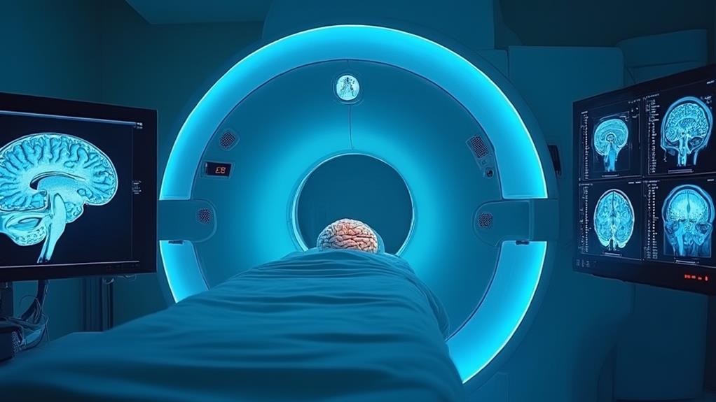
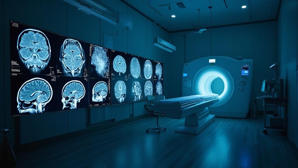
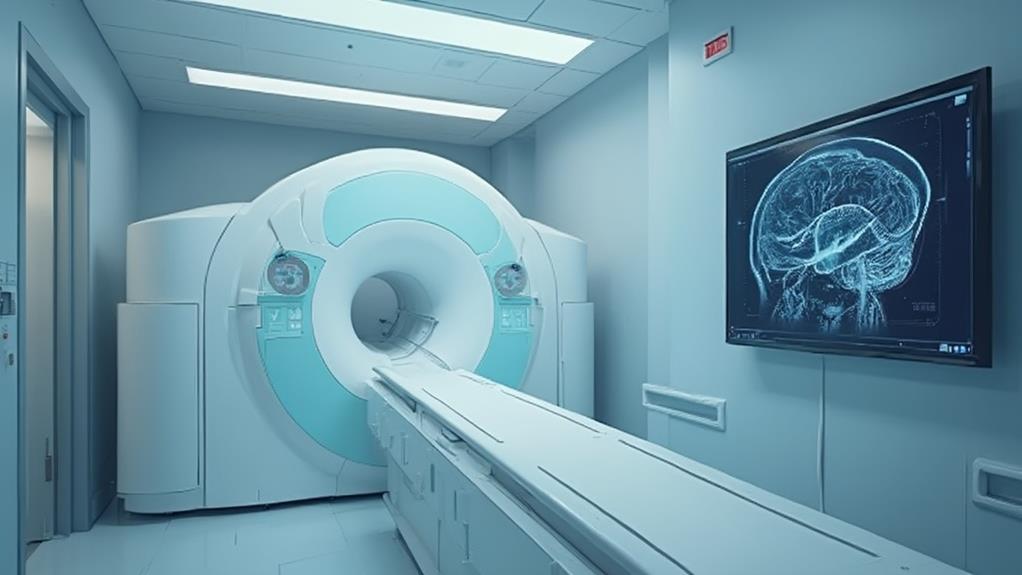
Magnetic Resonance Imaging (MRI) is an essential non-invasive tool for brain scans, generating detailed images of the brain's intricate structures. It employs magnetic fields and radiofrequency pulses to visualize soft tissues, offering superior contrast resolution compared to other imaging methods. MRI is instrumental in diagnosing neurological conditions such as tumors, strokes, multiple sclerosis, and neurodegenerative diseases. Its ability to differentiate between various tissues enables precise localization of abnormalities while avoiding ionizing radiation, making it safe for ongoing monitoring. Additionally, MRI provides critical, real-time information on brain function. Learn more about this technology's extensive applications and benefits.
MRI Highlights
- MRI scans provide high-resolution images of brain structures, aiding in the diagnosis of various neurological conditions.
- MRI uses a non-invasive approach, ensuring patient safety by eliminating the need for surgery or ionizing radiation.
- MRI excels in detecting minute abnormalities such as small tumors, microbleeds, and signs of neurodegenerative diseases.
- Functional MRI visualizes brain activity in real-time, useful for assessing brain functions and identifying affected regions.
- MRI is critical for early disease detection, enabling timely interventions and improving patient outcomes through continuous monitoring.
Definition and Usage Explained

Magnetic Resonance Imaging (MRI) provides detailed images of the brain, capturing the intricacies of brain structures with remarkable clarity. This non-invasive technique offers numerous benefits, including unparalleled soft tissue contrast, which facilitates the accurate diagnosis of various neurological conditions such as tumors, multiple sclerosis, and strokes.
MRI is especially valuable in brain scans for its capability to highlight soft tissue abnormalities, which makes it superior to other imaging methods for detecting subtle changes in brain tissue. The use of MRI in clinical practice is indispensable for its ability to reveal abnormalities invisible to other imaging methods, ensuring timely and effective treatment.
What MRI Scans Capture
MRI scans, an acronym for Magnetic Resonance Imaging, serve as a non-invasive diagnostic tool to visualize the brain's intricate structures. These scans capture high-resolution images that detail the brain's anatomy, enabling medical professionals to observe various regions and detect abnormalities. MRI technology employs strong magnetic fields and radiofrequency pulses to generate detailed images of the brain's internal composition, such as grey matter, white matter, and cerebrospinal fluid spaces.
The advanced capabilities of MRI scans allow for the detection of a range of neurological conditions. These include tumors, stroke damage, multiple sclerosis plaques, aneurysms, and traumatic brain injuries.
Additionally, MRI scans can provide critical information on brain development and integrity, instrumental in diagnosing developmental anomalies and neurodegenerative diseases like Alzheimer's.
Another remarkable aspect of MRI scans is their ability to capture functional information. Functional MRI (fMRI) measures brain activity by detecting changes in blood flow, reflecting neural activity. This capability is essential for mapping eloquent areas of the brain involved in critical functions such as movement, speech, and sensation, often aiding in pre-surgical planning. Through capturing these detailed and functional insights, MRI scans substantially enhance the understanding and treatment of numerous neurological conditions.
Benefits of Using MRI
In the domain of medical diagnostics, the advantages of utilizing MRI technology are multifaceted and significant. Magnetic Resonance Imaging (MRI) boasts a non-invasive approach, eliminating the need for surgery or the use of ionizing radiation. This guarantees patient safety while providing detailed images of the brain.
Additionally, MRI excels in differentiating between various tissues, such as gray and white matter, enhancing the precision of diagnoses.
The technology's superior contrast resolution makes it particularly effective for detecting minute abnormalities, including small tumors, microbleeds, and signs of neurodegenerative diseases. Functional MRI (fMRI) extends these capabilities by visualizing brain activity in real-time, aiding in the assessment of brain functions and detecting regions affected by disorders.
Moreover, MRI can be repeatedly used to monitor disease progression or therapeutic responses without cumulative radiation exposure, making it ideal for long-term patient management. Its adaptability also enables specialized scans, such as diffusion tensor imaging (DTI), which maps neural pathways, aiding in conditions like Multiple Sclerosis.
Common Brain Conditions Diagnosed
The superior imaging capabilities and safety profile of MRI technology have rendered it indispensable in diagnosing various brain conditions. MRI scans offer high-resolution images of brain structures, aiding physicians in identifying a multitude of neurological disorders. One of the most common conditions diagnosed through MRI is brain tumors. These scans can detect both benign and malignant growths, providing pivotal information on their size, type, and location, which guides treatment strategies.
MRI technology also excels in diagnosing multiple sclerosis (MS). By highlighting areas of demyelination, or damage to the myelin sheath, MRIs help in early detection and monitoring of MS progression. Strokes, both ischemic and hemorrhagic, are another critical area where MRI is instrumental. It allows for precise localization of affected brain regions, essential for timely and appropriate intervention.
Beyond physical anomalies, MRIs identify various types of neurodegenerative disorders such as Alzheimer's disease. They can reveal structural changes in brain anatomy, such as atrophy, which are indicative of these conditions. In addition, MRIs prove valuable in diagnosing epilepsy by identifying structural brain abnormalities that could be triggering seizures. This nondestructive, detailed imaging is indispensable for accurate diagnosis and effective management of these and many other brain conditions, ultimately enhancing patient care and outcomes.
Benefits

MRI for brain scans presents numerous advantages, beginning with its ability to assist in early disease detection, enabling timely medical interventions. The procedure is non-invasive, providing a safer alternative to more intrusive diagnostic methods.
Additionally, MRI technology produces high-resolution images, allowing for detailed brain mapping that can accurately pinpoint abnormalities and guide treatment plans. MRI is particularly useful for diagnosing conditions like tumors, hemorrhaging, and issues with the pituitary gland.
Early Disease Detection
Early detection of neurological diseases offers significant advantages in patient outcomes and overall healthcare management. Magnetic Resonance Imaging (MRI) plays a pivotal role in identifying abnormalities at an early stage, making it possible to intervene swiftly and effectively. This early intervention is crucial in preventing disease progression and enhances the potential for successful treatment.
Improved Treatment Outcomes: Early identification of conditions such as tumors, multiple sclerosis, or vascular disorders, allows for timely medical or surgical interventions, increasing the chances of recovery and improving quality of life.
Enhanced Monitoring: Routine MRI scans enable continuous monitoring of disease progression, allowing healthcare providers to adjust treatments based on real-time data, ensuring the best possible patient care.
Reduced Healthcare Costs: Early detection can mitigate the need for more extensive and expensive treatments required at later stages, contributing to overall cost-effectiveness within the healthcare system.
Better Prognosis: Detecting diseases before symptoms become severe often leads to a better prognosis, providing patients with more time to ponder and pursue a range of treatment options, enhancing both lifespan and health outcomes.
Early detection is a cornerstone of effective healthcare, and MRI is an indispensable tool in this vital process.
Non-Invasive Procedure
Neurological disease management greatly benefits from methods that identify conditions at an initial stage, fostering improved patient outcomes and reduced healthcare costs. One of the most significant advantages of MRI for brain scans is that it is a non-invasive procedure.
In contrast to other diagnostic methods that may require incisions or the insertion of instruments into the body, MRI employs strong magnetic fields and radio waves to generate detailed images of the brain without affecting any physical structures directly. This attribute makes MRI scans especially valuable for patient care.
Being non-invasive, MRI scans eliminate the risks associated with surgical procedures, such as infection or complications from anesthesia. Additionally, patients experience no discomfort during the process, which typically takes around 30 to 60 minutes, allowing them to remain calm and cooperative throughout. The absence of recovery time means that individuals can resume their daily activities almost immediately post-scan.
These qualities make MRI scans a safer and more comfortable alternative for patients, particularly those who may be vulnerable due to age or underlying health conditions. This non-invasive nature aligns well with the medical community's ongoing commitment to optimizing patient experience and minimizing unnecessary risks.
High-Resolution Images
In addition to its non-invasive nature, an important benefit of MRI for brain scans is its ability to produce high-resolution images. The clarity and detail of these images are paramount when diagnosing various neurological conditions, allowing healthcare professionals to provide more accurate and personalized care. The high-resolution capability greatly enhances the effectiveness of the diagnostic process.
Heightened Detection of Abnormalities: High-resolution MRI images enable the detection of small and subtle abnormalities that might be missed with lower-resolution scans, thereby facilitating early diagnosis and treatment.
Improved Diagnostic Precision: The detailed images assist in differentiating between various types of brain lesions, which is vital for accurate diagnosis and appropriate treatment planning.
Extensive View of Brain Structures: The ability to capture clear images of different brain regions allows for a more thorough assessment, aiding in the detection of a wide range of neurological disorders, from tumors to degenerative diseases.
Guided Interventions: High-resolution images provide precise guidance during neurosurgical procedures and other medical interventions, ensuring that treatments are targeted and minimizing damage to healthy tissue.
Detailed Brain Mapping
Detailed brain mapping through MRI offers numerous benefits, making it a cornerstone of advanced neurological diagnostics. One of the primary advantages is its non-invasive nature, which allows for an extensive understanding of brain structure and function without the need for surgery. This aspect is particularly crucial for patients who may be at risk from invasive procedures.
Furthermore, the high-resolution images provided by MRI enable clinicians to detect and monitor intricate abnormalities, such as small tumors, vascular malformations, or areas affected by neurodegenerative diseases. Early detection facilitated by detailed brain mapping can greatly improve patient outcomes by enabling prompt and precise interventions.
MRI also plays a pivotal role in pre-surgical planning. For patients undergoing brain surgery, such as those with epilepsy or brain tumors, detailed maps of the brain's functional areas help surgeons minimize damage to critical regions responsible for movement, speech, and other essential functions. This reduces the risk of postoperative complications and enhances the patient's quality of life.
Additionally, detailed brain mapping supports ongoing research efforts, contributing to the understanding of various neurological conditions. This research underpins the development of new treatments and therapeutic strategies, further demonstrating the multifaceted benefits of MRI in advancing neurological care and improving patient well-being.
Common Conditions Diagnosed

MRI scans are pivotal in diagnosing several critical brain conditions. Among the most common diagnoses are brain tumors, strokes, and multiple sclerosis, each of which presents distinct MRI features. The following table summarizes these conditions:
| Condition | Key MRI Findings |
|---|---|
| Brain Tumors | Abnormal growths, with or without contrast enhancement |
| Stroke and Damage | Areas of restricted blood flow, visible as bright spots or dark areas |
| Multiple Sclerosis | Lesions in the white matter, often seen as bright spots on T2-weighted images |
Detecting Brain Tumors
Brain tumors represent a significant and serious condition that MRI technology is adept at identifying. Magnetic Resonance Imaging (MRI) employs strong magnetic fields and radio waves to generate detailed images of brain structures, making it invaluable in detecting both benign and malignant tumors. The non-invasive nature of MRI, combined with its high-resolution capabilities, safeguards early and accurate diagnosis, which is pivotal for effective treatment planning and improving patient outcomes.
MRI scans excel in differentiating between various types of brain tumors. Four key ways in which MRI technology assists in detecting brain tumors:
- Structural Imaging: MRI provides clear images of brain anatomy, aiding in the identification of abnormal growths and lesion boundaries.
- Contrast Enhancement: Using gadolinium-based contrasts, MRI enhances visibility of tumor tissues, distinguishing them from healthy brain matter.
- Functional MRI (fMRI): This technique assesses brain activity by measuring changes in blood flow, helping pinpoint functional areas around a tumor for safer surgical planning.
- Advanced Imaging Techniques: Methods such as Diffusion Tensor Imaging (DTI) and Magnetic Resonance Spectroscopy (MRS) offer insights into tumor characterization and cellular composition, respectively.
Identifying Stroke and Damage
Strokes and neurological damage represent common conditions for which MRI technology serves as a pivotal diagnostic tool. MRI, or Magnetic Resonance Imaging, captures highly detailed images of the brain, allowing healthcare practitioners to identify areas affected by ischemia or hemorrhage. Immediate and accurate detection of strokes is essential in both minimizing harm and guiding appropriate treatment interventions.
Strokes result from interrupted blood flow to the brain, leading to potential loss of neurological function. MRI scans, particularly Diffusion-Weighted Imaging (DWI), are highly sensitive to acute ischemia. Within minutes of an ischemic stroke, DWI can identify affected brain tissue, thereby enabling rapid medical response. Additionally, MRIs help in detecting hemorrhagic strokes, characterized by bleeding within the brain, which require different treatment strategies compared to ischemic strokes.
Beyond stroke identification, MRI is instrumental in evaluating the extent of brain damage post-event. T2-weighted images and FLAIR sequences are particularly useful in detecting brain edema and long-term damage. These imaging techniques aid clinicians in planning rehabilitation and monitoring recovery progress, ensuring that interventions are tailored to the patient's specific neurological condition. Ultimately, MRI's role in diagnosing strokes and neurological damage is indispensable for effective, patient-centered care.
Diagnosing Multiple Sclerosis
When it comes to diagnosing multiple sclerosis (MS), an unpredictable and often debilitating condition, MRI technology is indispensable. MS involves the immune system attacking the myelin sheath that protects nerves, leading to a variety of neurological symptoms. MRI scans provide detailed images of the brain and spinal cord, which are critical for identifying the characteristic lesions associated with MS. These lesions, or plaques, appear as bright spots on the MRI scans, allowing for a more accurate diagnosis.
Understanding how MRI aids in diagnosing MS includes:
- Identifying Lesions: MRI is adept at highlighting areas of active inflammation and past damage in the brain and spinal cord.
- Monitoring Disease Progression: Through follow-up MRIs, clinicians can assess the evolution of MS, tracking the appearance of new lesions or changes in existing ones.
- Evaluating Treatment Efficacy: MRIs play a key role in measuring how well a patient is responding to treatment, guiding therapeutic decisions.
- Differentiating from Other Conditions: MRI helps differentiate MS from other neurological disorders that mimic its symptoms, such as neuromyelitis optica or acute disseminated encephalomyelitis.
Utilizing MRI in diagnosing MS allows healthcare professionals to deliver precise care, tailored to each individual's specific condition, fostering better outcomes for those affected by this challenging disease.
MRI FAQ
Is an MRI Scan Painful?
The current question is whether an MRI scan is painful. Rest assured, MRI scans are typically non-invasive and painless. Any discomfort usually arises from the need to remain still in an enclosed space for an extended period.
How Long Does an MRI Brain Scan Take?
The duration of a brain scan typically ranges from 20 to 45 minutes, depending on the complexity and the specific requirements of the examination. Efficient and empathetic patient care is critical to guarantee comfort during the procedure.
Can You Move During an MRI Scan?
Movement during a scan should be avoided to guarantee accurate results. Patients' cooperation is essential to obtaining high-quality images, and staying still minimizes the likelihood of needing repeat scans, thereby promoting efficient and effective diagnostic procedures.
Do I Need to Fast Before an MRI?
Regarding the current question, fasting is not typically required before an MRI unless instructed otherwise by your healthcare provider. Adhering to any specific guidelines guarantees the most accurate results and supports the care process.
Are There Any Risks Involved With MRI Brain Scans?
There are minimal risks involved with MRI brain scans. However, individuals with implanted medical devices like pacemakers must inform the technician. Additionally, some may experience mild anxiety or claustrophobia due to the enclosed space during the procedure.
