MRI contrast agents improve the clarity and diagnostic accuracy of magnetic resonance imaging by enhancing the visibility of tissues and abnormalities. They work by altering proton relaxation times, which increases the contrast between different tissues. These agents also facilitate detailed imaging of blood flow, vascular structures, and lesions that may otherwise be missed. Gadolinium-based agents are common, though manganese ions and iron oxide nanoparticles are also used. These agents can help in identifying tumors, inflammatory processes, and vascular anomalies, substantially aiding in accurate diagnoses and treatment planning. Understanding their variety and effectiveness can further enhance medical outcomes.
MRI Highlights
- MRI contrast agents help enhance image clarity and contrast, making it easier to distinguish between different types of tissues.
- Gadolinium-based agents improve proton relaxation times but pose risks for patients with kidney impairment.
- Manganese-based agents provide safer alternatives for tissue differentiation due to their endogenous nature and controlled dosing.
- Both gadolinium and manganese agents contribute to better detection of abnormalities like tumors, vascular lesions, and inflammatory processes.
- Contrast agents significantly enhance the contrast-to-noise ratio and signal intensity, providing detailed visualization of organ functionality and structure.
Purpose of MRI Contrast Agents

MRI contrast agents are utilized to enhance image clarity, allowing for better tissue differentiation and the detection of abnormalities within the body. By altering the local magnetic field, these agents improve the contrast between different types of tissues. This enhances the accuracy of diagnostic imaging, facilitating more precise evaluations and treatments.
Often, these agents are particularly beneficial for diagnosing abnormalities like tumors, hemorrhaging, and pituitary gland problems. By using a contrast agent, the technologist can obtain clearer and better diagnostic images, which is essential for planning the best course of treatment.
Enhancing Image Clarity
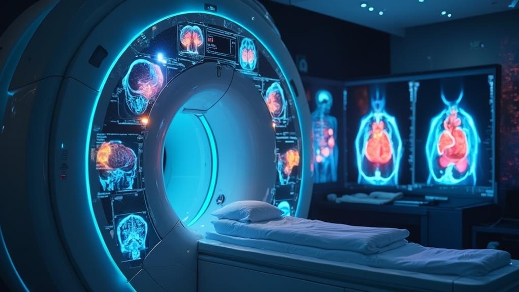
To achieve ideal diagnostic accuracy, medical practitioners often utilize contrast agents to enhance the clarity of magnetic resonance imaging (MRI) scans. These agents play an essential role not only in improving the visibility of internal structures but also in aiding healthcare professionals to render more accurate diagnoses.
Contrast agents often contain paramagnetic substances like gadolinium, which enhance the proton relaxation times in tissues. This results in a brighter and clearer image, helping in identifying subtle abnormalities.
By improving the visibility of blood flow and vascular structures, contrast agents allow for a detailed examination of blood vessels, which is pivotal for detecting issues such as aneurysms, blockages, or malformations.
Contrast agents can highlight lesions that might be missed in non-contrast scans. This is especially important in identifying and delineating tumors, infections, and inflammatory conditions.
Tissue Differentiation
Tissue differentiation is a pivotal function of MRI contrast agents, facilitating the clear distinction between different types of tissues within the body. By enhancing the contrast between various tissue types, these agents enable healthcare professionals to better visualize anatomical structures and assess physiological functions. This distinct visibility is essential in accurately analyzing complex regions where subtle differences in tissue composition may be imperceptible with standard MRI techniques.
The application of MRI contrast agents greatly improves the contrast-to-noise ratio, thereby augmenting the ability to identify and characterize tissues. For instance, contrast agents like gadolinium-based compounds interact with the magnetic field to enhance the relaxation rates of nearby hydrogen protons. This results in increased signal intensity from certain tissues, making them stand out more distinctly in the resultant images.
Furthermore, the ability to differentiate tissues accurately paves the way for more precise assessment of organ functionality and structure. It is particularly useful in evaluating soft tissues, which often present challenges in non-enhanced imaging due to their similar electromagnetic properties. Consequently, MRI contrast agents play an indispensable role in the thorough and precise evaluation of patients, directly impacting the effectiveness of diagnostic imaging. This dedication to precision ultimately supports medical professionals in delivering ideal patient care.
Detecting Abnormalities
While facilitating tissue differentiation enhances anatomical visualization, the primary objective of MRI contrast agents lies in their ability to detect abnormalities within the body. These agents improve image clarity by increasing the contrast between different tissues, making it easier to identify and characterize pathological conditions. The enhanced imaging capabilities help clinicians in diagnosing and monitoring various medical conditions.
MRI contrast agents are particularly valuable in spotting abnormalities due to the changes in vascular properties they highlight. Here are three specific ways these agents assist in detection:
- Tumor Identification: MRI contrast agents can help differentiate between benign and malignant tumors by enhancing the visibility of tumor vascularity and tissue heterogeneity.
- Inflammatory Processes: Contrast agents illuminate areas of inflammation, assisting in the identification and assessment of inflammatory diseases such as multiple sclerosis and rheumatoid arthritis.
- Vascular Abnormalities: They also make it possible to visualize blood vessels and identify vascular anomalies such as aneurysms, stenoses, and arteriovenous malformations.
These enhanced imaging capabilities aid healthcare providers in making accurate diagnoses, forming the basis for effective treatment strategies. By detecting abnormalities with precision, MRI contrast agents contribute to improved patient outcomes, aligning with the medical community's commitment to serving others.
Benefits

The utilization of MRI contrast agents provides significant benefits in medical imaging. These agents enhance image clarity, allowing for more detailed organ visualization and improved diagnostic accuracy.
Thanks to advances in high-resolution photos, contrast agents enable clinicians to make more precise diagnoses. In addition, they are instrumental in better tumor detection, contributing to more effective patient management and treatment planning.
Enhanced Image Clarity
Utilizing MRI contrast agents leads to markedly enhanced image clarity, which plays an essential role in accurate medical diagnostics. The efficacy of MRI scans is vastly improved by these agents, enabling healthcare providers to serve their patients with optimum accuracy and swiftness. Enhanced clarity allows medical professionals to make more precise assessments, reducing ambiguity in diagnosis and treatment planning.
MRI contrast agents contribute to enhanced image clarity in multiple ways:
- Improved Signal-to-Noise Ratio: By increasing the differentiation between distinct tissues, contrast agents help in highlighting abnormalities that might otherwise go unnoticed. This improvement empowers radiologists to identify conditions with a higher degree of confidence.
- Enhanced Detection of Pathologies: Detecting minute abnormalities, such as small tumors or vascular lesions, becomes more feasible. This accurate detection aids in timely intervention, which is critical in managing progressive diseases.
- Precision in Structural Detail: Fine details of anatomical structures become more pronounced, facilitating a deeper understanding of the patient's condition. This precision is invaluable in planning surgical procedures and other therapeutic interventions.
Detailed Organ Visualization
Delving deeper into the domain of MRI scans, detailed organ visualization stands out as a significant benefit bolstered by the use of MRI contrast agents. Contrast agents enhance the differentiation of tissues, making it possible to capture exquisite details of organs that might otherwise be indistinguishable. This level of detail is particularly advantageous in identifying subtle structural and functional abnormalities.
For medical professionals dedicated to optimizing patient care, this capability is indispensable. It allows the visualization of intricate anatomical details, including the vascular supply and fine tissue structures, thus facilitating a more extensive assessment of organ health. Such precision is vital when evaluating conditions like tumors, vascular diseases, and soft-tissue abnormalities.
Additionally, the improved visualization aids in planning surgical and therapeutic interventions by offering a clear and precise roadmap of the target organ, reducing the risk of complications. This enhances the overall effectiveness of treatment strategies, ensuring patients receive the most appropriate care tailored to their specific needs. For those in the healthcare field committed to serving others, the ability to achieve such fine-resolution images exemplifies a significant leap forward in diagnostic imaging technology. Ultimately, detailed organ visualization through MRI contrast agents plays a pivotal role in advancing patient outcomes.
Improved Diagnostic Accuracy
Harnessing the advanced capabilities of MRI contrast agents, improved diagnostic accuracy marks a transformative leap in medical imaging. The precision and clarity provided by these agents contribute significantly to accurate diagnosis and effective treatment planning, guaranteeing that patients receive the best possible care. By enhancing the visibility of internal structures and anomalies, MRI contrast agents empower healthcare providers to make more informed decisions.
Consider the following benefits that aid in achieving improved diagnostic accuracy:
- Enhanced Clarity: MRI contrast agents accentuate the differences between normal and abnormal tissues, leading to more definitive and clear images. This clarity is essential for identifying abnormalities that may otherwise go unnoticed.
- Detailed Imaging: These agents offer unparalleled detail, allowing for the visualization of intricate anatomical structures. Enhanced images mean that physicians can identify and diagnose conditions with a much higher degree of confidence.
- Accurate Assessments: With enhanced imaging capabilities, contrast agents enable more precise evaluations of disease progression and treatment efficacy, ensuring that therapeutic approaches can be finely tuned for ideal patient outcomes.
Such advancements in diagnostic accuracy ensure that medical professionals can serve their patients with a higher degree of precision and reliability, fostering improved health outcomes and enhanced patient care.
Better Tumor Detection
When it comes to tumor detection, MRI contrast agents present unparalleled benefits by vastly enhancing the capability to identify and characterize malignancies. By improving the visibility of abnormal tissue, these agents enable more precise differentiation between benign and malignant tumors. This heightened contrast allows for earlier detection and accurate staging, which is indispensable for effective treatment planning.
MRI contrast agents work by altering the magnetic properties of nearby hydrogen atoms in water molecules, which is particularly useful in highlighting vascularized tumor tissues. Enhanced imaging provides detailed insights into tumor size, shape, and location, facilitating more accurate biopsies. Additionally, these agents can differentiate between different types of tumors based on their vascularity and cellular environment, aiding in the customization of patient-specific treatment protocols.
Furthermore, utilizing MRI contrast agents minimizes the likelihood of false positives and negatives, thereby reducing unnecessary procedures and emotional strain on patients. Early and accurate detection not only aids in better prognosis but also impacts the overall effectiveness of therapeutic interventions. In this regard, MRI contrast agents play a pivotal role in advancing the quality of oncological care, ultimately contributing to improved patient outcomes and betterment of community health.
Types of MRI Contrast Agents

Understanding the types of MRI contrast agents is essential for enhancing image quality and ensuring accurate diagnoses. The main categories include Gadolinium-Based Contrast Agents, Iron Oxide Nanoparticles, and Manganese-Enhanced Imaging Options, each offering unique benefits and applications. The following table highlights key characteristics of these agents:
| Contrast Agent Type | Key Component | Primary Use |
|---|---|---|
| Gadolinium-Based Contrast Agents | Gadolinium | General Enhancement |
| Iron Oxide Nanoparticles | Iron Oxide | Liver and Spleen Imaging |
| Manganese-Enhanced Imaging Options | Manganese | Neuroimaging |
Gadolinium-Based Contrast Agents
Gadolinium-based contrast agents (GBCAs) substantially enhance the diagnostic capabilities of MRI by improving the visibility of internal tissues. These agents, indispensable in various medical diagnoses, leverage the properties of gadolinium—a rare earth metal with strong magnetic attributes. When administered to patients, GBCAs enable better differentiation between normal and abnormal tissues, assisting healthcare professionals in forming precise diagnoses and optimizing treatment plans.
Key benefits of GBCAs include:
- Enhanced Imaging: GBCAs provide clear images of blood vessels and the heart, pivotal for detecting blockages, aneurysms, or other vascular abnormalities.
- Tumor Detection: They help identify and delineate tumors, enabling precise characterization and aiding in the monitoring of tumor size and spread.
- Diagnosis of Inflammatory Conditions: GBCAs are instrumental in visualizing inflammatory processes, useful in conditions such as multiple sclerosis and inflammatory bowel disease.
Administering GBCAs must be done with care, as gadolinium is naturally toxic. To mitigate this, gadolinium is chelated—bound to carrier molecules—which minimizes risk and allows it to be safely excreted from the body. Overall, GBCAs greatly elevate the diagnostic value of MRI, thereby profoundly impacting patient care and outcomes through more accurate and earlier diagnosis.
Iron Oxide Nanoparticles
Among the alternative MRI contrast agents, iron oxide nanoparticles (IONPs) present a promising option, owing to their unique magnetic properties and biocompatibility. These nanoparticles enhance signal contrast by shortening T2 relaxation times in tissues, thereby creating darker images in T2-weighted MRI scans. Their magnetic cores, typically composed of magnetite (Fe3O4) or maghemite (γ-Fe2O3), attribute these properties, while their biocompatible coatings facilitate stability in biological systems.
IONPs exhibit high sensitivity for detecting pathologies, particularly in liver and spleen imaging, due to their propensity for uptake by the mononuclear phagocyte system. This makes them instrumental in identifying metastatic liver lesions with high spatial resolution. Moreover, their surface can be functionalized with various ligands, enabling targeted imaging of specific tissues or biomarkers, which greatly aids in the early detection of diseases.
Importantly, iron, being a naturally occurring element in the human body, reduces the risk of long-term toxicity, which is a notable advantage over some conventional contrast agents. By promoting precision in diagnostic imaging, IONPs support clinicians in providing more accurate diagnoses and tailored treatments, ultimately enhancing patient care and outcomes. Their continued development heralds a new era in MRI technology, with profound implications for medical diagnostics.
Manganese-Enhanced Imaging Options
As an alternative to traditional MRI contrast agents, manganese-enhanced imaging options leverage the paramagnetic properties of manganese ions (Mn2+) to enhance image contrast. Manganese ions interact with water protons, resulting in the shortening of T1 relaxation times, which provides a brighter image in T1-weighted MRI scans. This capability makes manganese-enhanced imaging a compelling tool for various clinical applications.
The following are the key features and benefits of manganese-enhanced imaging options:
- Neuroimaging Applications: Manganese ions can traverse neuronal pathways, aiding in the mapping of neural networks and providing detailed insights into brain function and architecture. This property is particularly useful for studying conditions such as neurodegenerative diseases.
- Cellular-level Imaging: Manganese ions can penetrate cell membranes and enhance intracellular contrast, making them valuable for detailed cellular and molecular imaging. This allows researchers to observe cell processes and interactions more clearly.
- Safety Profile: Manganese is an endogenous element, and when used in controlled doses, it poses fewer risks compared to gadolinium-based agents, which have been associated with nephrogenic systemic fibrosis in patients with kidney impairment.
Incorporating manganese-enhanced imaging in clinical practice could support more accurate diagnostics and better patient outcomes, aligning with the mission to serve others by improving healthcare delivery.
MRI FAQ
Are MRI Contrast Agents Safe for Children and Pregnant Women?
Evaluating the safety for children and pregnant women requires careful consideration. Pediatric and maternal health must always be prioritized, ensuring that any diagnostic procedure, including the use of contrast agents, is warranted and conducted with utmost safety protocols.
Can MRI Contrast Agents Cause Allergic Reactions?
Allergic reactions, while generally rare, can occur. Ensuring patient safety requires vigilance and readiness to manage potential reactions. Providers must assess each individual's risk factors, ensuring informed decisions and compassionate care for all patients.
How Long Does an MRI With Contrast Take Compared to One Without?
An MRI with contrast typically requires an additional 15-30 minutes to administer the contrast agent and perform enhanced imaging. In comparison, a standard MRI generally lasts between 30-60 minutes, depending on the complexity and area examined.
Are There Any Dietary Restrictions Before an MRI With Contrast?
Typically, there are no dietary restrictions before undergoing this procedure. However, fasting might sometimes be recommended. It's best to follow specific instructions provided by healthcare professionals to guarantee the most accurate and safe imaging results.
What Should I Do if I Feel Side Effects After an MRI With Contrast?
If side effects occur after the procedure, it is prudent to contact your healthcare provider immediately. Describe the symptoms in detail, and follow their guidance to facilitate prompt and appropriate care for your well-being.
