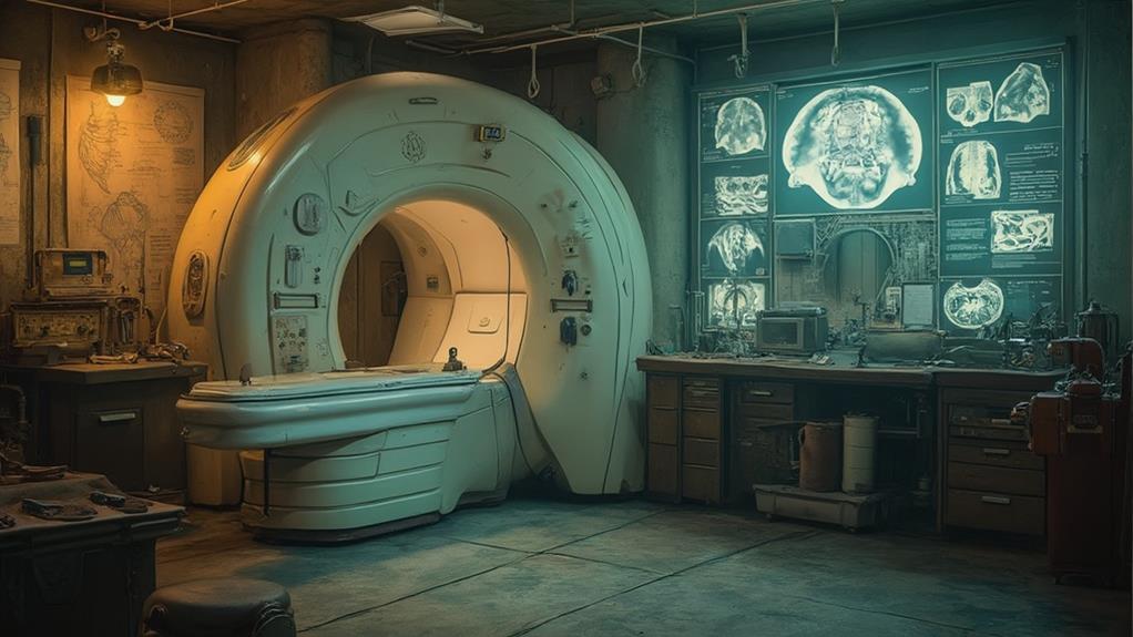
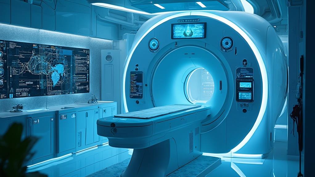
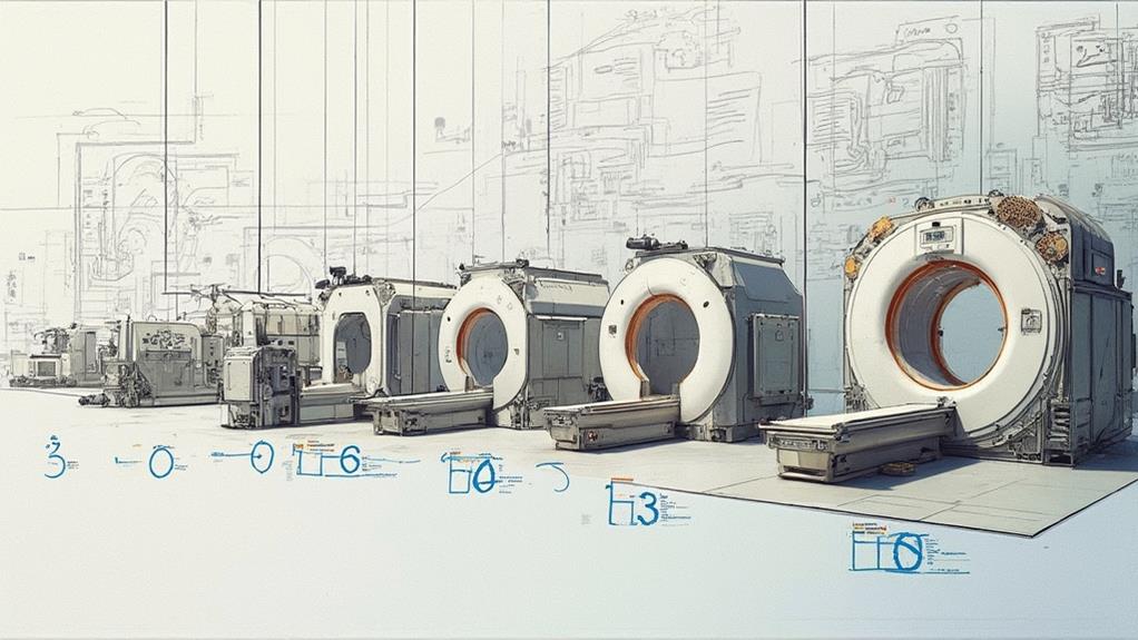
The history of MRI technology began in the early 1970s with significant breakthroughs in research. Raymond Damadian discovered that different tissues and tumors exhibited varying relaxation times in a magnetic field, demonstrating NMR's potential for medical imaging. Paul Lauterbur further advanced this by introducing magnetic field gradients, enabling two-dimensional imaging. Peter Mansfield refined the technology, leading to the first clinical MRI systems in the 1980s, which offered unprecedented insights into soft tissue contrast. Early applications in neurology and orthopedics showcased MRI's non-invasive diagnostic capabilities, marking a milestone in healthcare. Key advancements and collaborations have since optimized this indispensable medical tool. Continue exploring to uncover further technological progress and clinical applications.
MRI Highlights
- Pioneering efforts in MRI began in the early 1970s with Damadian's discovery of varying relaxation times in tissues.
- Lauterbur introduced magnetic field gradients for 2D imaging, transforming NMR from spectroscopy to imaging.
- Mansfield refined the technology with echo-planar imaging, speeding up image acquisition.
- Initial clinical applications in the 1980s provided critical insights into neurology, orthopedics, and cardiac functions.
- MRI offers detailed soft tissue imaging without radiation, improving early disease detection and patient safety.
Invention of MRI Technology

The inception of MRI technology was marked by pivotal early research breakthroughs that laid the groundwork for its development, with key contributions from inventors such as Paul Lauterbur and Sir Peter Mansfield. Their pioneering work in the 1970s led to the refinement of techniques that enabled the generation of detailed images of the human body's internal structures.
These advancements soon translated into initial clinical applications, revolutionizing medical diagnostics and establishing MRI as an indispensable tool in modern healthcare. Early MRI machines provided superior diagnostic images, which substantially helped in evaluating symptoms like vertigo, headaches, seizures, or vision issues.
Early Research Breakthroughs
Pioneering efforts in the field of MRI technology can be traced back to the early 1970s, when researchers like Raymond Damadian and Paul Lauterbur made significant strides. During this period, the scientific community was exploring the potential of nuclear magnetic resonance (NMR) for medical imaging.
Damadian's critical work demonstrated that the relaxation times of different tissues and tumors varied when placed in a magnetic field, laying foundational theories for MRI. This discovery suggested that NMR could be employed to distinguish between healthy and diseased tissue.
In parallel, Paul Lauterbur introduced a groundbreaking concept that revolutionized imaging techniques. He proposed the use of magnetic field gradients to create two-dimensional images. Lauterbur's innovative approach allowed for the spatial localization of NMR signals, effectively transforming NMR from a mere spectroscopy tool into a powerful imaging modality – what we now know as MRI (Magnetic Resonance Imaging). These early breakthroughs established MRI's core principles, enabling subsequent technological advancements.
This nascent period was marked by rapid advancements and collaborative research, ultimately setting the stage for the clinical MRI systems that would emerge in the ensuing decades. This transforming contribution underscored the commitment to medical advancement and the betterment of patient care.
Key Inventors' Contributions
Several pivotal figures contributed to the invention of MRI technology, greatly shaping its development and application. Among these, Raymond Damadian is often recognized for his early work in the 1970s that demonstrated the potential for nuclear magnetic resonance (NMR) to differentiate between normal and cancerous tissue. This laid the groundwork for MRI technology.
Paul Lauterbur advanced this field considerably by introducing the concept of creating two-dimensional images using magnetic field gradients. This innovation enabled the conversion of NMR data into spatially informative images, effectively birthing what we now know as MRI.
Peter Mansfield further refined MRI technology through his pioneering work on echo-planar imaging (EPI). His advancements allowed for the rapid acquisition of detailed images, enhancing the practicality and efficiency of MRI in medical diagnostics.
Lastly, the collective efforts of numerous researchers and engineers worldwide have continuously optimized MRI systems, improving their resolution, speed, and safety. The collaborative nature of this technological evolution highlights a shared commitment to advancing medical imaging for better patient outcomes. These key contributions underscore the importance of interdisciplinary collaboration in advancing healthcare technologies, ensuring that patients receive the highest standard of diagnostic care.
Initial Clinical Applications
With the foundational work of key inventors laying the groundwork, MRI technology soon found its way into clinical settings. The 1980s marked the dawn of initial clinical applications, underscoring the growing potential for non-invasive diagnostic imaging. The first systems, though rudimentary by today's standards, provided unprecedented insights into soft tissue contrast.
Physicians were swift to adopt this technology for its ability to differentiate between normal and abnormal tissues with remarkable clarity. Neurology and orthopedics were among the earliest fields to benefit, utilizing MRI to visualize brain tumors, spinal cord abnormalities, and joint disorders. This significant advancement in imaging facilitated more accurate diagnoses and improved patient outcomes.
Moreover, the versatility of MRI soon became apparent. Beyond static images, dynamic studies of blood flow and cardiac function emerged, expanding the clinical applications further. MRI's ability to provide detailed anatomical and functional information without the risks associated with ionizing radiation resonated strongly within the medical community. This emphasis on patient safety and improved diagnostic precision marked a significant milestone.
In essence, the initial clinical applications of MRI technology underscored its transformative potential, laying a robust foundation for ongoing advancements. The medical community's embrace of MRI mirrored their unwavering commitment to enhancing patient care and diagnostic accuracy.
Benefits

MRI technology offers a range of significant benefits, making it an invaluable tool in modern medicine. It provides detailed images of soft tissues without exposing patients to radiation, ensuring a safer diagnostic process.
Additionally, MRI systems, like those in affordable MRI services, excel in patient comfort by accommodating individuals with mobility challenges and reducing anxiety through high-field, open designs. Its ability to detect diseases at an early stage greatly enhances treatment outcomes.
Non-invasive Diagnostic Tool
How has medical imaging evolved to provide accurate diagnoses without intrusive procedures? Medical Resonance Imaging (MRI) technology represents a significant leap in non-invasive diagnostic tools. These advancements allow healthcare professionals to examine internal structures without the need for surgical procedures, reducing patient risk and discomfort.
MRI, in particular, uses powerful magnets and radio waves to create detailed images of the body, providing clinicians with vital information for diagnosis and treatment.
The non-invasive nature of MRI technology offers several benefits:
- Reduced Patient Risk and Discomfort: Unlike invasive procedures, MRI does not involve incisions or exposure to ionizing radiation, minimizing risks such as infection and prolonged recovery times.
- Swift and Accurate Diagnoses: The high-quality images produced by MRI enable precise assessments of various conditions, leading to quicker and more precise diagnoses, which is essential for timely treatment.
- Versatility in Diagnosing Multiple Conditions: MRI can be used to diagnose a wide range of medical conditions, from neurological disorders to musculoskeletal injuries, making it a versatile tool in a healthcare provider's arsenal.
Detailed Soft Tissue Images
Delving deeper into the advantages of MRI technology, its ability to produce detailed soft tissue images stands out as a paramount benefit. This capacity allows healthcare professionals to obtain high-resolution images of tissues such as the brain, muscles, and organs, which are essential for accurate diagnosis and treatment planning. By utilizing powerful magnets and radio waves, MRI technology surpasses other imaging modalities in capturing the nuances of soft tissue structures.
For instance, the clarity of MRI images aids in the thorough examination of neurological conditions, facilitating early detection of brain tumors or multiple sclerosis. This precision not only enhances diagnostic accuracy but also improves patient outcomes by enabling timely and targeted interventions.
Additionally, MRI is invaluable in evaluating muscle injuries and ligament tears, providing vital information without invasive procedures. Moreover, the versatility of MRI allows it to differentiate between various tissue types, offering a more extensive view of complex anatomical regions. This capability is particularly beneficial in oncology, where distinguishing between malignant and benign tissues can greatly influence treatment strategies.
Hence, the detailed soft tissue imaging provided by MRI technology is instrumental in advancing medical care, aiding professionals dedicated to serving and improving patient health.
No Radiation Exposure
Beyond the ability to capture intricate soft tissue details, another pivotal advantage of MRI technology is its non-reliance on radiation. This characteristic inherently enhances patient safety, especially when multiple imaging sessions are required. Unlike CT scans and X-rays, MRI does not utilize ionizing radiation, making it a preferable option for various patient groups, including pediatric populations and those requiring frequent imaging.
MRI's non-reliance on radiation brings several essential benefits:
- Patient Safety: The absence of ionizing radiation eliminates the risk of radiation-induced complications, thus reducing the potential for long-term adverse effects. This is especially beneficial for vulnerable groups such as pregnant women and children.
- Repetitive Imaging: Patients with chronic conditions or those undergoing follow-up examinations can safely undergo multiple MRI scans without the cumulative risks associated with repeated radiation exposure, ensuring continuity in monitoring and diagnostics.
- Broader Applicability: MRI's safe profile broadens its applicability across diverse patient demographics, including those with pre-existing health conditions that might be exacerbated by radiation, thereby enabling extensive and inclusive healthcare services.
This attribute of MRI technology underscores its indispensable role in modern diagnostic imaging, contributing considerably to patient well-being and the overall effectiveness of healthcare provision.
Early Disease Detection
Early disease detection stands as one of the most vital benefits afforded by MRI technology. Utilizing magnetic resonance imaging, healthcare professionals can identify abnormalities in tissues and organs at their earliest stages, often before symptoms become apparent. This capability is essential for diseases such as cancer, where early intervention greatly improves prognosis and survival rates. For instance, small tumors in the brain, spine, or other critical areas can be identified and monitored with precision, allowing for timely medical intervention.
Moreover, MRI technology plays a pivotal role in detecting conditions affecting the brain, such as Multiple Sclerosis (MS), where early diagnosis can lead to better management and improved quality of life. Its ability to produce detailed, clear images without the use of ionizing radiation makes MRI a preferred method for long-term monitoring of chronic conditions, ensuring patient safety over extended periods.
The advancement of MRI technology underscores its commitment to patient-first healthcare, empowering medical providers with the tools needed to detect and treat ailments promptly. This not only enhances individual patient outcomes but also contributes to reducing the broader societal burden of disease, aligning with the healthcare community's mission of service and well-being for all.
Further Advancements in MRI

Building on the fundamental principles of MRI, further advancements have considerably enhanced imaging resolution, enabled the development of functional MRI (fMRI), and introduced hybrid imaging techniques. These innovations allow for more precise and detailed visualization of anatomical structures, functional assessments of brain activity, and the integration of multiple imaging modalities. The following table summarizes key advancements, their impact, and applications:
| Advancements | Impact | Applications |
|---|---|---|
| Enhanced Imaging Resolution | Improved anatomical detail | Neuroimaging, oncology |
| Functional MRI Development | Real-time brain activity mapping | Cognitive neuroscience, clinical research |
| Hybrid Imaging Techniques | Combined functional and anatomical data | Personalized medicine, multifaceted diagnosis |
| Advanced Sequence Design | Rapid imaging protocols | Emergency diagnostics, pediatric imaging |
| AI Integration | Enhanced data analysis | Predictive diagnostics, treatment planning |
Enhanced Imaging Resolution
The pursuit of precision has driven significant advancements in MRI technology, particularly in enhancing imaging resolution. High-resolution imaging has become a cornerstone of MRI, allowing for unprecedented detail in visualizing internal structures. This progress is important for serving the medical community by providing clearer, more accurate diagnostics.
Progress in enhanced imaging resolution can be summarized in the following key areas:
- Increased Magnetic Field Strength: Innovations in MRI technology have led to the use of higher magnetic field strengths, such as 3T (Tesla) and even 7T. Higher field strengths improve image quality by yielding better signal-to-noise ratios, which are vital for capturing minute anatomical details.
- Advanced Gradient Coils: The development of stronger and more precise gradient coils has directly contributed to increased spatial resolution. These coils generate rapid, accurate changes in the magnetic field, leading to finer image slices and superior image detail.
- Improved RF Coils: Radiofrequency (RF) coils used in MRI machines have also seen enhancements. Modern coils are designed to be more sensitive and capable of capturing detailed images from specific regions with greater accuracy, aiding in focused diagnostics.
Functional MRI Development
Alongside the strides made in enhancing imaging resolution, functional MRI (fMRI) has emerged as a pivotal advancement in the field. Unlike traditional MRI, which focuses on structural imaging, fMRI tracks brain activity by measuring changes in blood flow. This non-invasive technique relies on detecting fluctuations in oxygen levels within blood, reflecting the brain's activity. When neurons in a specific brain region activate, local blood flow increases, bringing oxygen-rich blood that alters the magnetic properties of surrounding tissues.
Developed in the early 1990s, fMRI has revolutionized our understanding of the human brain. Researchers and clinicians employ fMRI to observe cognitive functions, map sensory processing, and pinpoint regions implicated in various neurological and psychiatric disorders. By offering real-time insights into brain network dynamics, fMRI promotes a better grasp of previously elusive neural mechanisms.
The versatility of fMRI also extends to clinical applications, aiding in pre-surgical planning for patients undergoing brain surgery. By mapping essential functional areas, fMRI assists neurosurgeons in making informed decisions, ensuring the preservation of key cognitive functions. Continual advancements in fMRI technology promise even greater precision and utility, fostering improved diagnostic and therapeutic strategies in neurology and psychiatry.
Hybrid Imaging Techniques
Pushing the boundaries of imaging capabilities, hybrid imaging techniques signify a pivotal leap forward in MRI technology. These advancements merge the strengths of different imaging modalities, such as MRI combined with Positron Emission Tomography (PET) or Computed Tomography (CT), consequently enhancing diagnostic and therapeutic strategies.
Hybrid imaging offers several key benefits:
- Augmented Diagnostic Accuracy: By combining anatomical and functional imaging, hybrid systems provide more extensive and precise information. PET/MRI, for example, integrates metabolic function with high-resolution tissue structure, aiding in more accurate diagnoses.
- Improved Patient Care: The integration of multiple imaging modalities reduces the need for patients to undergo separate scans, leading to quicker diagnostics and treatment plans, ultimately resulting in more efficient patient management and care.
- Advanced Research Potential: These techniques open new avenues for clinical research. They allow for the simultaneous evaluation of structure and function, providing deeper insights into diseases and potentially leading to the discovery of novel biomarkers.
Hybrid imaging techniques not only advance the field of radiology but also improve patient outcomes and foster innovations in medical research. They represent the continuous evolution of MRI technology, driven by the commitment to enhance medical diagnostics and treatments for the benefit of patients.
MRI FAQ
What Safety Concerns Should Be Considered Before Undergoing an MRI Scan?
Before undergoing an MRI scan, safety concerns include ensuring the absence of metal implants or devices, avoiding the scan during pregnancy unless essential, and disclosing medical conditions like kidney issues that might interfere with contrast agents.
How Long Does a Typical MRI Scan Take?
A typical MRI scan usually takes between 30 to 60 minutes. However, the exact duration can vary depending on the specific area being examined and the complexity of the procedure, ensuring precise and high-quality diagnostic results.
Can All Types of Medical Implants Be Scanned Using MRI Technology?
No, not all medical implants are compatible with MRI technology. It is essential to assess each patient's implant specifics to avoid risks, ensuring the scan proceeds safely and without compromising the integrity of the implant.
How Has the Cost of MRI Scans Changed Over the Years?
The cost of MRI scans has generally increased over the years due to technological advancements, increased demand, and enhanced diagnostic capabilities. Efforts to make MRI more accessible and affordable continue to be a priority in the healthcare industry.
Are There Any Alternatives to MRI for Imaging Soft Tissues?
Yes, alternatives to MRI for imaging soft tissues include ultrasound and computed tomography (CT) scans. Ultrasound offers real-time imaging, while CT provides detailed cross-sectional images. Both methods serve distinct diagnostic purposes in patient care and medical practice.
