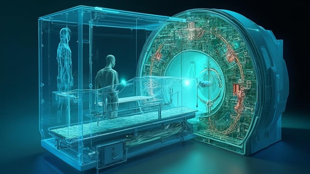
There are several prevalent misconceptions about MRI that warrant clarification. Primarily, MRI is not painful and does not involve invasive procedures or the use of ionizing radiation. Instead, it uses magnetic fields and radio waves, making it a safer alternative to X-rays and CT scans, even for pregnant women and children. Another common myth is that MRI is unsafe for those with metal implants, but safety precautions are in place to manage this. Ultimately, while some may find the procedure uncomfortable due to noise and enclosed space, it remains a highly valuable diagnostic tool offering detailed internal images. Understanding these aspects can help demystify the MRI experience.
MRI Highlights
- MRI scans are not painful and do not require anesthesia or sedation.
- MRI does not use ionizing radiation, making it safer than X-rays and CT scans.
- MRI is safe for frequent use, including during pregnancy and for children.
- MRI machines are designed to accommodate mild discomfort from enclosed spaces, with technicians monitoring patients.
- Specific safety precautions are necessary, including screening for metal implants and removing personal items containing metal.
How MRI Works

To understand how MRI works, it is essential to grasp the basics of magnetic fields, which play a pivotal role in creating detailed body images. The imaging process involves aligning hydrogen atoms using a strong magnetic field, then capturing the emitted signals as the atoms return to their normal state.
This process is helpful in evaluating a variety of symptoms such as vertigo, headaches, and seizures. MRI scans are particularly beneficial because they do not require any radiation, making it a safer option for obtaining high-quality images. Safety protocols and precautions are rigorously followed to guarantee patient and technician safety throughout the procedure.
Magnetic Field Basics
Magnetic Resonance Imaging (MRI) leverages the principles of physics to produce detailed images of the body's internal structures. Central to this technology is the utilization of a powerful magnetic field, alongside radiofrequency pulses, to align hydrogen atoms within the body.
When placed in the strong magnetic field, protons within these hydrogen atoms align with the direction of the field. This magnetic alignment is fundamental to MRI technology. Once aligned, radiofrequency pulses are applied, knocking the protons out of alignment. These protons then return to their original state, a process called relaxation. During relaxation, they emit energy signals that are detected by the MRI scanner's coils.
The time it takes for the protons to realign, as well as the amount of energy they release, varies depending on the type of tissue they are in—such as muscle, fat, or bone.
Imaging Process Explained
Understanding the intricate imaging process of MRI discloses a fascinating interplay between technology and human anatomy. At its core, MRI utilizes powerful magnetic fields and radio waves to generate highly detailed images of the internal structures of the body.
The process begins with the patient lying inside a large, cylindrical magnet. Inside this magnet, a strong and consistent magnetic field aligns the protons in the body's hydrogen atoms.
Radiofrequency pulses are then sent through the body, causing these aligned protons to produce a faint signal. After the radio frequency pulse is turned off, the protons return to their original alignment, emitting energy in the form of radio waves as they do so. These emitted signals are captured by receiver coils and translated into digital images by sophisticated computer algorithms.
The contrasts in the images arise from differences in the density of hydrogen atoms as well as the varying relaxation times of the protons in different tissues. This differentiation allows clinicians to pinpoint problems with an exceptional level of clarity, emphasizing MRI's critical role in diagnostic medicine. The non-invasive nature of MRI guarantees a highly detailed analysis without the need for surgical procedures, making it an invaluable tool for patient care.
Safety and Precautions
Guaranteeing patient safety during an MRI procedure involves adhering to a few indispensable precautions due to the powerful magnetic field and radiofrequency pulses inherent in the technology. First and foremost, patients must be thoroughly screened for any metallic implants or devices, such as pacemakers, cochlear implants, or metal clips, which can interfere with the MRI's magnetic field and potentially cause harm. Personal items containing metal, including jewelry and watches, must also be removed before the scan.
Another important precaution involves communication. Patients should inform their healthcare provider of any medical conditions, such as kidney issues, as the contrast agents sometimes used in MRI scans can affect renal function. For individuals with claustrophobia, medication or sedation may be arranged to guarantee a comfortable experience during the often enclosed scanning process.
Staff training is equally vital. Technicians and radiologists must be well-versed in operating the MRI machine and recognizing potential contraindications. Additionally, clear guidelines should be maintained for emergency procedures to address any complications swiftly.
Lastly, continuous monitoring is essential. Observing patients throughout the MRI process helps in detecting any adverse reactions early, guaranteeing swift and effective responses, thereby optimizing patient safety and comfort.
Benefits

MRI offers a multitude of benefits, making it a valuable diagnostic tool in modern medicine. This imaging method is pain-free, provides detailed views of internal structures, and is non-invasive, allowing for safe and thorough assessments.
Unlike other imaging techniques, MRI requires no radiation, making it a safer option for many patients. It is particularly useful for diagnosing abnormalities like tumors and hemorrhaging, ensuring accurate and effective diagnoses of a wide range of medical conditions.
These attributes contribute to its frequent use in diagnosing a wide range of medical conditions accurately and effectively.
Pain-free Imaging Method
Although medical procedures often carry an inherent fear of pain or discomfort, Magnetic Resonance Imaging (MRI) stands out as a pain-free diagnostic method that offers numerous benefits. Unlike some medical interventions that may involve invasive techniques or cause physical discomfort, MRI scans are non-invasive and typically painless. Patients lie still inside the MRI machine, remaining comfortable while the machine generates detailed images of internal structures.
One significant advantage of MRI is the elimination of exposure to ionizing radiation, unlike X-rays or CT scans. This feature is particularly beneficial for patients requiring frequent monitoring or for those with conditions necessitating multiple imaging sessions. Additionally, the lack of radiation makes MRI a safer option for vulnerable populations, such as pregnant women and children, who might be more susceptible to the harmful effects of radiation.
Furthermore, MRI procedures are generally stress-free and do not require anesthesia, sedation, or any recovery period. This convenience allows individuals to resume their daily activities immediately after the scan, making it an efficient diagnostic tool. For healthcare professionals committed to patient care, MRI provides a reliable, non-disruptive, and safe method to obtain critical information, thereby enhancing the overall quality and effectiveness of medical diagnostics.
Detailed Internal Views
With its unparalleled ability to produce detailed internal views, MRI technology stands at the forefront of modern diagnostic tools. It excels in capturing high-resolution images of the body's internal structures, providing an invaluable advantage for healthcare professionals striving to deliver ideal patient care. This technology leverages strong magnetic fields and radio waves to generate comprehensive images, often revealing conditions that may not be visible through other imaging methods.
Detailed internal views obtained from MRIs allow clinicians to accurately diagnose a broad range of medical conditions. These include, but are not limited to, abnormalities in the brain and spinal cord, joint disorders, and cancers. The precision of MRI images assists healthcare providers in devising targeted treatment plans, thus boosting the effectiveness of the interventions.
Furthermore, MRI's ability to deliver three-dimensional views ensures that the smallest details are discerned, aiding in the early detection of diseases. This early detection is indispensable for achieving better prognoses, enabling prompt and possibly less invasive treatments. Physicians and healthcare workers can rely on MRI imaging to exhaustively understand their patients' conditions and serve them with the highest degree of accuracy and care.
Non-invasive Procedure Option
One significant advantage of MRI technology is that it offers a non-invasive procedure option, minimizing patient discomfort and eliminating the need for surgical intervention. Unlike procedures that require incisions, MRI uses powerful magnetic fields and radio waves to create detailed images of the body's internal structures. This means there is no need for anesthesia or recovery time, making the process more convenient for patients.
The non-invasive nature of MRI is particularly beneficial for patients with underlying health conditions. It reduces the potential risks associated with surgery, such as infections or complications from anesthesia. Additionally, it allows for the repeated imaging of patients who require ongoing monitoring, such as those with chronic diseases, without subjecting them to additional physical stress.
Moreover, MRIs are well-suited for pediatric patients and the elderly, who may be more susceptible to the risks of invasive procedures. The ability to provide extensive diagnostic information without surgical intervention exemplifies the profound impact of MRI technology on patient care. This reduces overall healthcare costs by avoiding the expenses associated with invasive surgeries and ensuring quicker, simpler recovery times for patients, thereby enhancing their overall well-being and quality of life.
Safe Diagnostic Technique
Beyond its non-invasive nature, MRI technology stands out as an exceptionally safe diagnostic technique, offering numerous benefits that set it apart in the domain of medical imaging. Initially, MRI does not use ionizing radiation, which is commonly associated with potential health risks in other imaging modalities like X-rays and CT scans. This lack of radiation minimizes the risk of long-term side effects and makes MRI a safer option, especially for vulnerable populations such as pregnant women and children.
Moreover, MRI provides unparalleled contrast between different types of soft tissues, significantly enhancing the ability to diagnose a wide variety of conditions without the need for invasive procedures. This detailed imaging capability supports early and accurate diagnosis, which is imperative for the timely initiation of treatment plans that can lead to better patient outcomes.
In addition, the MRI technique allows for repeated imaging over time without corresponding cumulative risk, a critical advantage for monitoring chronic conditions and evaluating ongoing treatment efficacy. Additionally, advancements in MRI technology have led to improvements in patient comfort, such as quieter machines and shorter scan times, making the experience less challenging for patients. These attributes collectively underscore MRI's standing as a highly beneficial and safe diagnostic tool for modern medicine.
Safety Concerns and Considerations

When considering MRI safety, it is vital to address common misconceptions about radiation exposure, metal implant risks, and the impacts of noise and claustrophobia on patients. Despite popular beliefs, MRI scans do not utilize ionizing radiation, making them safer compared to other imaging techniques. The table below summarizes important considerations for each safety aspect:
| Safety Concern | Key Points |
|---|---|
| Radiation Exposure | MRI uses magnetic fields, not ionizing radiation. |
| Metal Implants | Specific implants can be scanned, but precautions are necessary. |
| Noise Levels | MRIs can be loud, but ear protection is provided. |
| Claustrophobia | Open MRI machines and sedation are available options. |
| Patient Screening | Detailed screening is essential to confirm safety. |
Radiation Exposure Myths
A common misconception about Magnetic Resonance Imaging (MRI) is that it involves exposure to harmful radiation. This belief likely stems from the confusion between MRI and other imaging techniques, such as X-rays and CT scans, which do indeed use ionizing radiation. Unlike these methods, MRI utilizes strong magnetic fields and radiofrequency waves to generate detailed images of structures within the body. This fundamental difference means that MRI does not expose patients to ionizing radiation, thereby enormously reducing potential health risks associated with radiation exposure.
MRI provides a safe, non-invasive means to inspect soft tissues, organs, and other internal structures. It is especially valuable for diagnosing conditions in the brain, spinal cord, muscles, and joints. Patients and healthcare providers alike can take comfort in the knowledge that MRI technology prioritizes patient safety by eliminating the radiation risks inherent in other imaging modalities. This understanding is essential for making informed decisions about imaging options and promoting ideal patient outcomes.
Metal Implant Risks
Traversing the domain of MRI scans requires comprehending specific safety considerations, particularly concerning individuals with metal implants. Metal objects can interact with the strong magnetic fields used in MRI, presenting potential risks and complications. Misunderstanding these risks can create undue anxiety or dismiss pivotal safety protocols.
Key considerations include:
- Magnetic Field Interference: Some metal implants, such as certain pacemakers or aneurysm clips, can be affected by MRI's strong magnetic fields, potentially leading to malfunction or displacement.
- Heating: Metal objects can heat up during an MRI scan, due to interaction with the electromagnetic field, raising concerns about tissue damage.
- Image Distortion: Metal implants can cause artifacts or distortions in MRI images, which may obscure diagnostic information, leading to incomplete assessments.
- Material Compatibility: Not all metals pose the same risk; titanium and non-ferromagnetic materials are generally MRI-safe, whereas ferromagnetic materials may pose significant hazards.
Patients with metal implants should always consult their healthcare provider before undergoing an MRI. Detailed assessments and alternative imaging methods may be considered to guarantee safety and accurate diagnosis. Healthcare professionals are dedicated to individual well-being, and prioritizing detailed evaluations fosters safer, more effective patient care.
Noise and Claustrophobia
Experiencing an MRI scan can be challenging for some patients due to noise and enclosed spaces. The noise generated by the machine's powerful magnets can be loud and repetitive, often leading to discomfort. This can be mitigated by providing patients with earplugs or headphones that play music, reducing the impact of the sound. It's essential for healthcare providers to ensure that patients are well-prepared for these auditory experiences, facilitating a smoother scanning process.
Claustrophobia is another common concern. The MRI scanner's design can make patients feel enclosed, potentially triggering anxiety. Open MRI machines, which have wider openings and provide more space, can be an alternative for those severely affected by claustrophobia. Comfort measures, such as calming techniques or mild sedatives, can also aid in alleviating anxiety.
Addressing these considerations demonstrates a commitment to patient-centered care. By offering options and preparing patients, medical professionals can substantially enhance their MRI experience. For those working in healthcare, being attentive to these aspects helps build a supportive environment, which is fundamental in delivering prime patient care. It is vital to acknowledge and address these concerns, promoting a more compassionate and effective healthcare experience.
MRI FAQ
Can I Eat Before an MRI Scan?
Yes, you can generally eat before an MRI scan. However, certain types of MRI procedures, like those involving the abdomen, may require fasting. Always confirm specific instructions with your healthcare provider to guarantee accurate results.
How Long Does an MRI Scan Typically Take?
An MRI scan typically takes between 30 to 60 minutes, depending on the area being examined and the specific diagnostic requirements. Patients' cooperation during this time contributes considerably to the accuracy and efficiency of the imaging process.
Is It Normal to Feel Claustrophobic During an MRI?
Experiencing claustrophobia during an MRI is not uncommon. Patients often feel confined within the scanner. Healthcare professionals can offer calming techniques and support to guarantee the experience is as comfortable and stress-free as possible.
Can I Listen to Music During the Procedure?
Yes, patients can often listen to music during an MRI procedure. This option not only helps provide a comforting and more enjoyable experience but also assists in reducing anxiety for those undergoing this diagnostic examination.
Do I Need a Referral for an MRI?
Yes, generally a referral from a physician is required for an MRI. This guarantees that the procedure is medically necessary and allows the imaging center to coordinate accurate diagnostic needs for ideal patient care and service.
