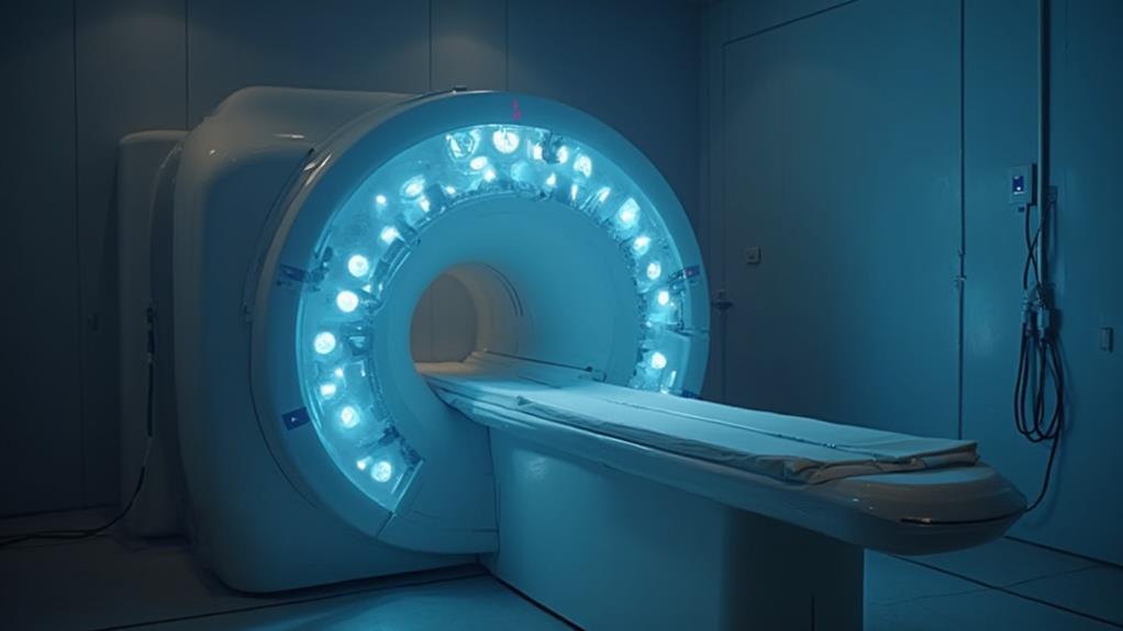
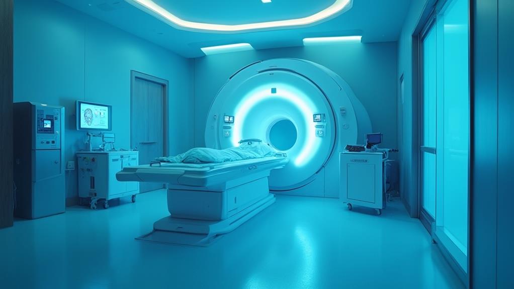
MRI scans, while critical for accurate medical diagnosis, come with certain risks. Exposure to strong magnetic fields can lead to projectile accidents and potential burns from metallic implants. Contrast agents, particularly gadolinium-based ones, may cause nephrogenic systemic fibrosis in individuals with kidney issues and allergic reactions. Claustrophobia and anxiety are common, and the loud acoustic noise generated by the machine can cause hearing damage if proper ear protection is not used. Temporary side effects include headaches, dizziness, and nausea. For a thorough understanding of these risks, more detailed information is essential.
MRI Highlights
- Strong magnetic fields pose risks for patients with metallic implants or devices.
- Gadolinium-based contrast agents can cause severe reactions, including nephrogenic systemic fibrosis, especially in patients with kidney disease.
- Acoustic noise from MRI scanners can lead to temporary or permanent hearing damage; ear protection is required.
- Patients often experience anxiety or claustrophobia during MRI scans due to the confined space and loud noises.
- Tissue heating from electromagnetic fields can cause discomfort, especially in areas with metal implants or electronic devices.
MRI Scan Definition

Magnetic Resonance Imaging (MRI) is a non-invasive diagnostic technique that uses a powerful magnetic field and radio waves to produce detailed images of the body's internal structures. The principle behind MRI involves aligning hydrogen atoms in the body's water and fat molecules, then detecting the energy released as they return to their original positions.
This imaging process allows for high-resolution visuals of soft tissues, muscles, and organs, essential in medical diagnoses and treatment planning. MRI is especially beneficial as it requires no radiation and provides safe, excellent images for evaluating symptoms like vertigo, headaches, and seizures. This method is indispensable for diagnosing abnormalities such as tumors, hemorrhaging, and pituitary gland problems.
MRI Basics Explained
An MRI scan, or Magnetic Resonance Imaging scan, is a non-invasive diagnostic tool that uses powerful magnets and radio waves to create detailed images of the internal structures of the body. This sophisticated technology allows clinicians to diagnose and monitor a variety of medical conditions by providing high-resolution images without the need for surgical interventions.
The principle advantage of an MRI scan is its ability to produce clear, detailed images of soft tissues, which can often be a challenge for other imaging methods such as X-rays or CT scans. This detail is particularly useful in examining the brain, spinal cord, joints, and organs like the heart and liver. MRI scans can identify abnormalities such as tumors, inflammation, or tissue damage, aiding in prompt and accurate treatment.
- Precision in Diagnosis: Enables clinicians to detect diseases in their early stages.
- Non-invasive Technique: Offers a pain-free method for obtaining critical diagnostic information.
- Versatility: Suitable for imaging a wide range of bodily tissues and conditions.
In the context of patient care, the ability of MRI to offer meticulous and clear images guarantees that healthcare providers can proceed with confidence, advocating for the well-being of those they serve.
Magnetic Field Principles
Understanding the principles behind MRI scans necessitates delving into the intricacies of magnetic field interactions. MRI, or Magnetic Resonance Imaging, employs strong magnetic fields and radio waves to generate images of the body's internal structures. Central to this technology is the behavior of hydrogen protons within the body, predominantly found in water and fat molecules.
In a typical MRI scan, a powerful magnet creates a stable, uniform magnetic field. This field aligns the magnetic moments of hydrogen protons in a specific direction. When radiofrequency (RF) pulses are applied, these protons are momentarily displaced from their alignment. As the RF pulse ceases, protons return to their original state, releasing energy in the process. This emitted energy is then detected by the MRI scanner, which converts it into detailed images.
The strength of the magnetic field, usually measured in Teslas (T), considerably impacts image clarity. Diagnostic MRI machines commonly operate within 1.5T to 3T. Understanding these magnetic field principles is critical for medical professionals in ensuring excellent imaging while upholding patient safety. This comprehension also enhances one's ability to communicate potential risks associated with MRI scans, underscoring the importance of informed decision-making in healthcare.
Imaging Process Overview
Having explored the principles behind the creation of magnetic fields, it is important to define the specifics of the imaging process in an MRI scan. An MRI, or Magnetic Resonance Imaging, leverages magnetic fields and radio waves to produce detailed images of the internal structures of the body. Unlike X-rays or CT scans, MRI does not involve ionizing radiation, making it a preferred choice for detailed imaging, particularly of soft tissues.
The process begins by positioning the patient inside a large tube-shaped magnet. When inside the scanner, radio waves are transmitted to create a resonance effect with hydrogen atoms in the body. These atoms emit signals that are captured by detectors and processed by a computer to form images. The resulting high-resolution images allow medical professionals to diagnose a wide range of conditions.
- Concern for patient comfort: During an MRI scan, the need for patients to remain still can be challenging and stressful, especially for those with claustrophobia.
- Dependence on advanced technology: The sophisticated nature of MRI machines means they are costly and predominantly chiefly available in well-equipped medical facilities.
- Time-consuming procedures: MRI scans can be lengthy, sometimes taking up to an hour, requiring patient patience and endurance.
Benefits

Magnetic Resonance Imaging (MRI) scans offer significant benefits, primarily improving diagnostic accuracy by providing detailed images of the body's internal structures. This non-invasive procedure allows for early disease detection, potentially leading to better patient outcomes.
The high-resolution imaging quality of MRI enhances the ability to identify and monitor various medical conditions effectively. MRI is especially useful for diagnosing abnormalities such as tumors, hemorrhaging, and problems with the pituitary gland. Essentially, it requires no radiation, making it a safer option for repeated imaging.
Improved Diagnostic Accuracy
One of the primary benefits of MRI scans is their exceptional diagnostic accuracy, which has revolutionized the field of medical imaging. This diagnostic tool leverages magnetic fields and radio waves to produce detailed images of the body's internal structures, greatly enhancing the ability to identify and diagnose a wide range of medical conditions. The precision of MRI scans has several profound impacts on patient care, enabling earlier and more accurate diagnosis of diseases, which can lead to better outcomes.
Caregivers and medical professionals will find the following emotional benefits compelling:
- Confidence in Diagnosis: The detailed images provided by MRIs guarantee that diagnoses are more accurate, which can lead to more effective treatment plans and peace of mind for patients.
- Early Detection: The high resolution of MRI scans allows for the early detection of medical conditions, particularly in areas such as oncology, neurology, and cardiology, thereby improving the likelihood of successful treatment.
- Informed Decision-Making: MRI scans offer thorough insights that can guide doctors and surgeons in making informed decisions about the best course of action, ultimately enhancing patient care.
In essence, the improved diagnostic accuracy of MRI scans empowers healthcare providers to deliver superior medical care, fulfilling their mission to serve and heal.
Non-Invasive Procedure
MRI scans provide a substantial advantage in the domain of non-invasive diagnostic procedures, offering a safer and more comfortable alternative to more invasive methods. By eliminating the need for surgical intervention, MRI technology enables healthcare professionals to obtain detailed images of internal structures without the associated risks and recovery time of traditional surgery. This aspect greatly benefits patients who are unable or unwilling to undergo more invasive diagnostic techniques due to underlying health conditions or personal preferences.
Furthermore, MRI scans involve no exposure to ionizing radiation, which is a critical safety feature, particularly for pregnant women and individuals requiring frequent imaging. The non-invasive nature of MRI enhances patient comfort, reducing anxiety and the physical strain associated with more intensive diagnostic procedures. This makes MRI an ideal choice for patients across various demographics.
The advanced imaging capabilities provided by MRIs also allow for the detection and monitoring of various medical conditions with high precision, aiding in timely and effective clinical decision-making. The seamless integration of MRI technology into routine clinical practice underscores its pivotal role in modern healthcare, ensuring that patient welfare remains a top priority.
Early Disease Detection
Early detection of diseases substantially benefits from the advanced imaging capabilities of MRI scans. By utilizing magnetic resonance technology, MRI provides detailed images of internal structures, allowing physicians to identify early signs of various conditions accurately. This early detection is essential in initiating timely interventions which can greatly improve patient outcomes and prevent the progression of diseases.
MRI scans are particularly valuable in detecting:
- Tumorous growths: Early identification of tumors, benign or malignant, facilitates prompt treatment strategies tailored to the specific type and stage of cancer, thereby increasing survival rates.
- Neurological disorders: Conditions such as multiple sclerosis, aneurysms, and strokes can be diagnosed in their nascent stages, enabling early management which can mitigate long-term damage and improve quality of life.
- Cardiovascular diseases: MRI can detect abnormalities in the heart and blood vessels early on, allowing for early intervention to prevent serious cardiac events such as heart attacks and strokes.
For healthcare providers dedicated to serving others, the capability of MRI to detect diseases at an early stage is invaluable. It not only enhances the chances of treatment success but also alleviates the emotional and physical burden on patients by addressing health concerns before they become critical.
Detailed Imaging Quality
The superior imaging quality provided by MRI scans presents substantial benefits in medical diagnostics. Unlike other imaging modalities, MRI offers an unparalleled ability to differentiate between various soft tissues within the body. This distinction is especially pivotal for diagnosing conditions involving complex and delicate structures, such as the brain, spinal cord, joints, and internal organs.
The level of detail attainable with MRI scans allows healthcare professionals to visualize abnormalities that might be obscured or undetectable with alternative imaging techniques, like X-ray or CT scans. For instance, subtle variations in soft tissue contrast enable the identification of lesions, tumors, and areas of inflammation with greater accuracy. This level of detail is essential for developing precise treatment plans, potentially improving patient outcomes, and reducing the likelihood of repeat diagnostic procedures.
Furthermore, the non-invasive nature of MRI scans means that high-quality images can be obtained without exposing patients to ionizing radiation. This characteristic is particularly advantageous for individuals requiring multiple scans over time, such as those with chronic conditions. In essence, the advanced imaging quality of MRI scans is a significant benefit, enhancing diagnostic precision and ultimately aiding in the provision of high-quality patient care.
Potential Side Effects Overview

| Issue | Severity | Description |
|---|---|---|
| Tissue Heating | Mild to Moderate | May cause slight warmth in scanned area |
| Temporary Discomfort | Mild | Feelings of claustrophobia or loud noises |
| Contrast Agent Reactions | Mild to Severe | Allergic reactions to gadolinium-based dyes |
| Skin Burns | Rare | From metallic objects left on the body |
| Visual Disturbances | Rare and Temporary | Blurred vision during or after the procedure |
Tissue Heating Concerns
When undergoing MRI scans, tissue heating stands as a significant concern due to the interaction between the electromagnetic fields and the body's tissues. This phenomenon occurs because the radiofrequency energy used in MRI can cause tissues, especially those with higher electrical conductivity, to absorb energy, leading to a rise in temperature. The extent of heating can vary depending on several factors including the duration of the scan, the strength of the magnetic field, and the specific absorption rate (SAR) of the energy.
Healthcare providers need to be aware of and monitor the potential for tissue heating, as excessive heating can lead to discomfort or even damage. Key considerations include:
- Patient discomfort: Heating may cause patients to feel warmth or a burning sensation in specific areas, making the experience uncomfortable.
- Vulnerable tissues: Certain tissues such as the eyes and testes are more susceptible to heating and may require additional caution.
- Implant concerns: Patients with metal implants or electronic devices such as pacemakers may face heightened risks, as these materials can exacerbate tissue heating.
Temporary Discomfort Explained
Temporary discomfort during and after an MRI scan is a common experience for many patients and typically manifests in several mild side effects. One of the most frequently reported sensations is a feeling of claustrophobia, primarily due to the enclosed nature of the MRI machine. Patients may find the confined space uncomfortable, which can sometimes lead to anxiety. Healthcare providers often suggest techniques such as deep breathing or visualizing calm environments to manage these feelings.
Additionally, the loud noises generated by the MRI machine, which include thumping and tapping sounds, can be unsettling. Providers routinely offer earplugs or headphones with music to mitigate the auditory discomfort. Another typical issue is lying still for extended periods, which can cause mild muscle stiffness or soreness. This effect tends to subsist shortly after the conclusion of the scan.
Moreover, the cold temperature within the MRI suite, necessary to operate the MRI's superconducting magnets, can cause minor physical discomfort. Blankets are usually provided to help keep patients warm. Ultimately, some patients report a slight dizziness or lightheadedness upon standing after the procedure, a temporary sensation that generally fades quickly. Overall, these side effects are manageable and temporary, ensuring the benefits of obtaining critical diagnostic information outweigh the discomfort.
Contrast Agent Reactions
Contrast agents, frequently used during MRI scans to enhance image clarity, can occasionally trigger adverse reactions in some patients. These agents, primarily gadolinium-based, are essential for detailed imaging but are not without risks. While many patients experience no issues, some may confront side effects ranging from mild to severe.
Common mild reactions include nausea, headache, and dizziness. Some individuals may exhibit allergic-like responses, causing rashes, itching, or shortness of breath. Though rare, severe reactions such as anaphylactic shock and nephrogenic systemic fibrosis (NSF) may occur, particularly in those with pre-existing kidney issues.
Patients with kidney problems are at heightened risk since gadolinium can accumulate, leading to NSF, a condition that causes skin thickening and organ dysfunction. Ensuring awareness and appropriate pre-screening are indispensable in mitigating these risks.
- Personal stories of families impacted by side effects may foster empathy.
- The commitment of healthcare professionals to patient safety reassures trust.
- Ongoing research and improvements reflect dedication to better care.
In light of these potential issues, it is necessary for healthcare providers to carefully screen for risk factors, inform patients about possible side effects, and promptly address any adverse reactions to uphold the highest standards of patient care.
MRI FAQ
Can I Wear Jewelry During an MRI Scan?
No, you should not wear jewelry during an MRI scan. Metal objects can interfere with the imaging process and may pose safety hazards. Ensuring patient safety and accurate results is our top priority.
How Long Does an MRI Scan Typically Take?
An MRI scan typically takes between 30 to 60 minutes, varying based on the complexity and area being studied. Duration guarantees thorough imaging, enabling better diagnostics to aid healthcare professionals in providing exemplary patient care.
Is It Safe to Have an MRI if I Am Pregnant?
Yes, it is generally safe to have an MRI while pregnant. However, it is essential to inform your healthcare provider about your pregnancy to guarantee appropriate precautions are taken to protect both you and your baby.
What Should I Do if I Feel Claustrophobic in the MRI Machine?
If you experience claustrophobia in the MRI machine, notify the technician immediately. They can provide solutions such as sedation, open MRI options, or other accommodations to guarantee your comfort while serving your needs effectively.
How Should I Prepare for an MRI Scan?
To prepare for an MRI scan, guarantee you inform the staff of any implants or medical conditions, remove all metal objects, and wear comfortable clothing. Arriving early can aid in relaxation and enhance service efficiency.
